

Development of novel adenosine monophosphate-activated protein kinase activators
- PMID: 20170185
- PMCID: PMC2841718
- DOI: 10.1021/jm901773d
Development of novel adenosine monophosphate-activated protein kinase activators
Retraction in
-
Retraction of "Development of Novel Adenosine Monophosphate-Activated Protein Kinase Activators".J Med Chem. 2018 Jun 14;61(11):5055. doi: 10.1021/acs.jmedchem.8b00707. Epub 2018 May 18. J Med Chem. 2018. PMID: 29772897 Free PMC article. No abstract available.
Abstract
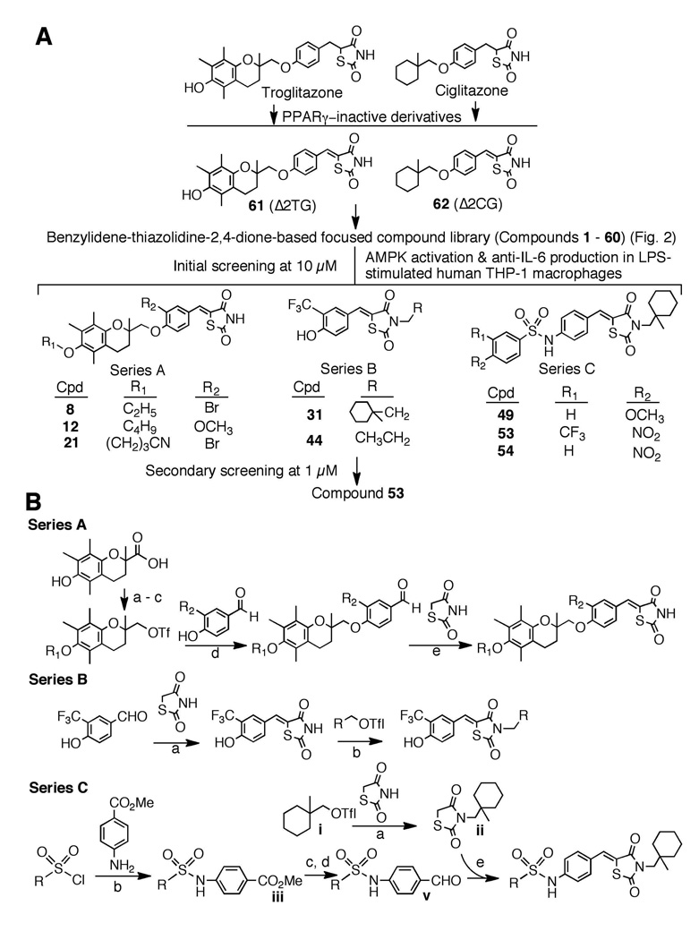
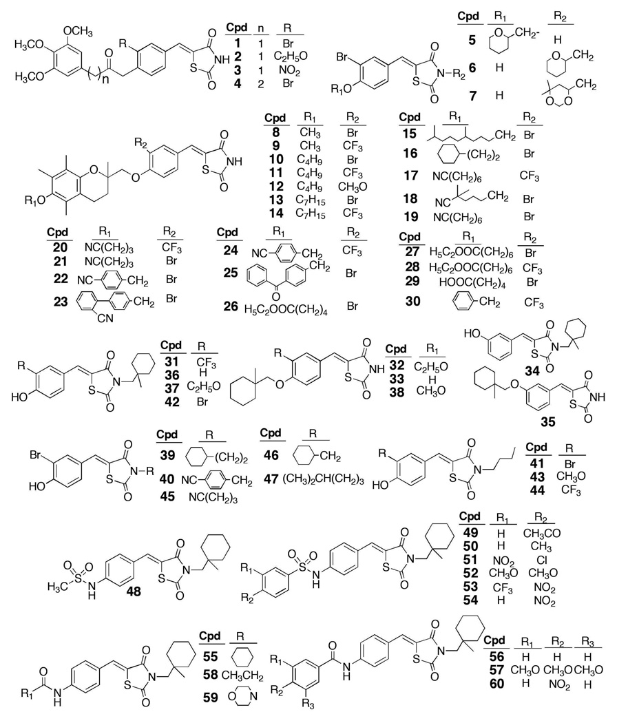
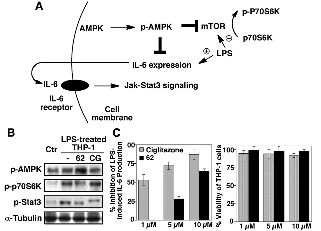
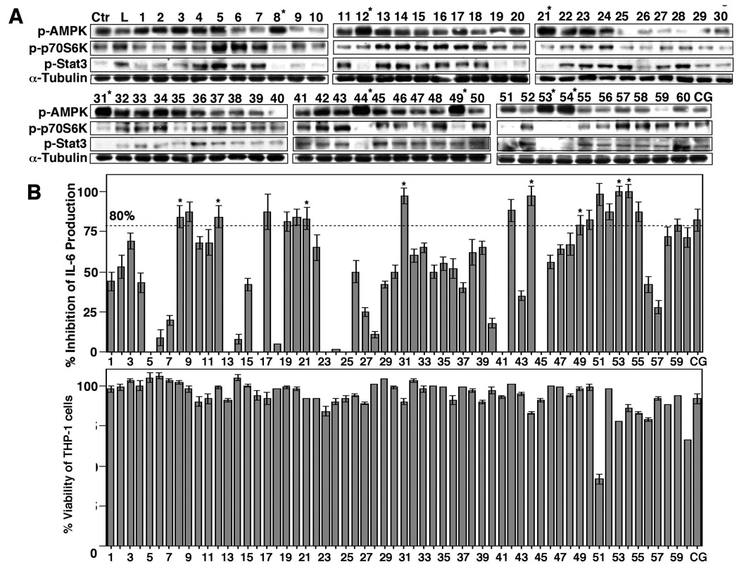
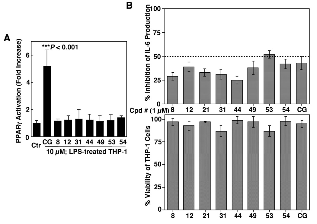
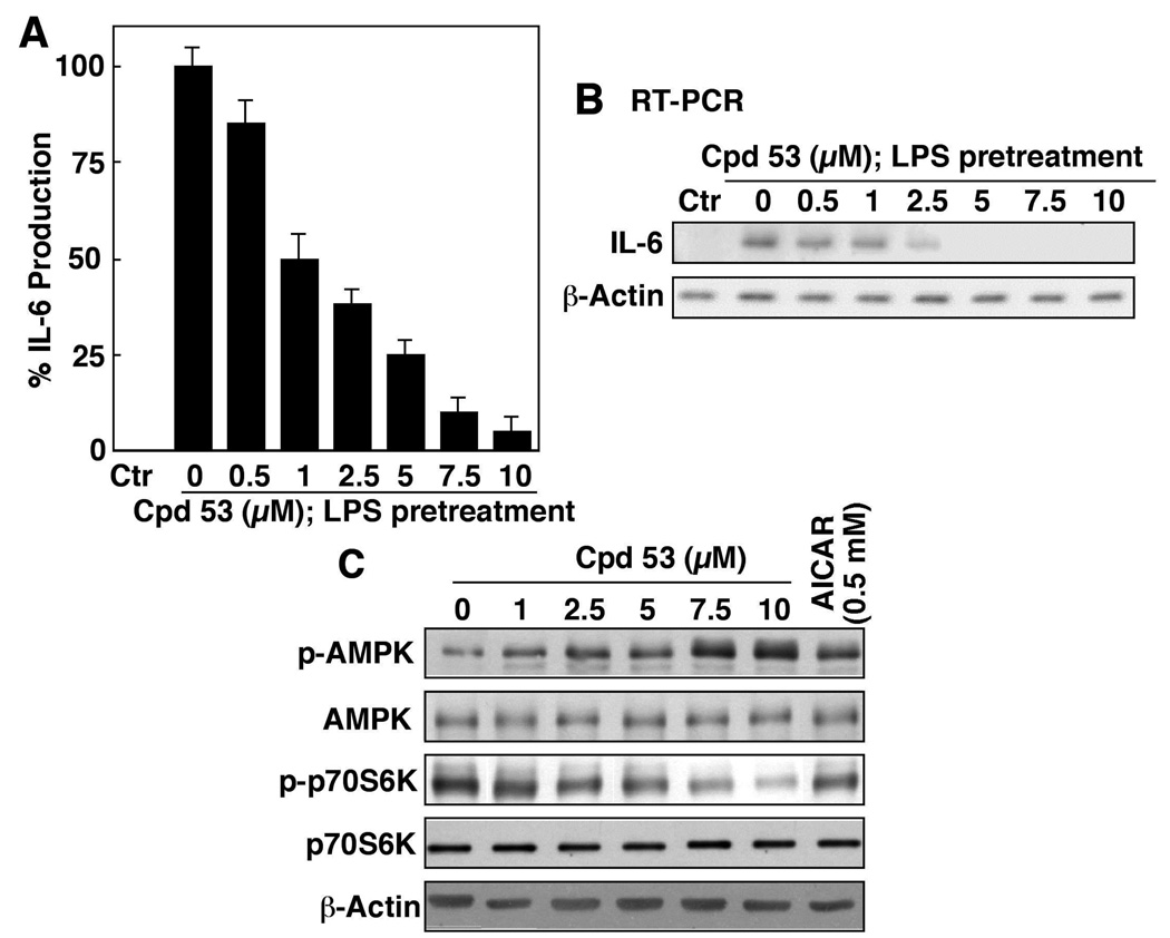
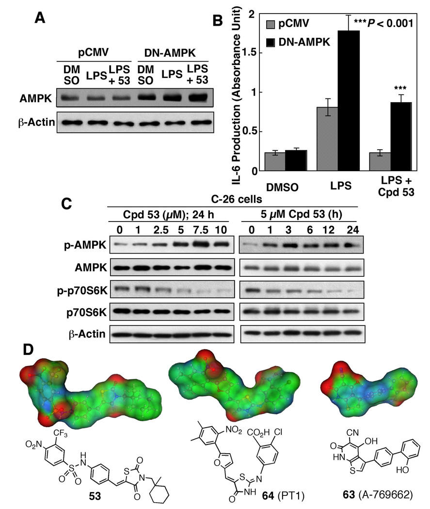
In light of the unique ability of thiazolidinediones to mediate peroxisome proliferator-activated receptor (PPAR)gamma-independent activation of adenosine monophosphate-activated protein kinase (AMPK) and suppression of interleukin (IL)-6 production, we conducted a screening of an in-house, thiazolidinedione-based focused compound library to identify novel agents with these dual pharmacological activities. Cell-based assays pertinent to the activation status of AMPK and mammalian homologue of target of rapamycin (i.e., phosphorylation of AMPK and p70 ribosomal protein S6 kinase, respectively) and IL-6/IL-6 receptor signaling (i.e., IL-6 production and signal transducer and activator of transcription 3 phosphorylation, respectively) in lipopolysaccharide (LPS)-stimulated THP-1 human macrophages were used to screen this compound library, which led to the identification of compound 53 (N-{4-[3-(1-methyl-cyclohexylmethyl)-2,4-dioxo-thiazolidin-5-ylidene-methyl]-phenyl}-4-nitro-3-trifluoro-methyl-benzenesulfonamide) as the lead agent. Evidence indicates that this drug-induced suppression of LPS-stimulated IL-6 production was attributable to AMPK activation. Furthermore, compound 53-mediated AMPK activation was demonstrated in C-26 colon adenocarcinoma cells, indicating that it is not a cell line-specific event.
Figures
Similar articles
-
Targeting energy metabolic and oncogenic signaling pathways in triple-negative breast cancer by a novel adenosine monophosphate-activated protein kinase (AMPK) activator.J Biol Chem. 2011 Nov 11;286(45):39247-58. doi: 10.1074/jbc.M111.264598. Epub 2011 Sep 14. J Biol Chem. 2011. PMID: 21917926 Free PMC article.
-
Development of Potent Adenosine Monophosphate Activated Protein Kinase (AMPK) Activators.ChemMedChem. 2015 Nov;10(11):1915-23. doi: 10.1002/cmdc.201500371. Epub 2015 Sep 9. ChemMedChem. 2015. PMID: 26350292 Free PMC article.
-
Small-molecule activators of AMP-activated protein kinase (AMPK), RSVA314 and RSVA405, inhibit adipogenesis.Mol Med. 2011 Sep-Oct;17(9-10):1022-30. doi: 10.2119/molmed.2011.00163. Epub 2011 Jun 1. Mol Med. 2011. PMID: 21647536 Free PMC article.
-
Recent progress in the identification of adenosine monophosphate-activated protein kinase (AMPK) activators.Bioorg Med Chem Lett. 2016 Nov 1;26(21):5139-5148. doi: 10.1016/j.bmcl.2016.09.065. Epub 2016 Sep 28. Bioorg Med Chem Lett. 2016. PMID: 27727125 Review.
-
Natural activators of adenosine 5'-monophosphate (AMP)-activated protein kinase (AMPK) and their pharmacological activities.Food Chem Toxicol. 2018 Dec;122:69-79. doi: 10.1016/j.fct.2018.09.079. Epub 2018 Oct 2. Food Chem Toxicol. 2018. PMID: 30290216 Review.
Cited by
-
Targeting myeloid-derived suppressor cells using a novel adenosine monophosphate-activated protein kinase (AMPK) activator.Oncoimmunology. 2016 Jul 25;5(9):e1214787. doi: 10.1080/2162402X.2016.1214787. eCollection 2016. Oncoimmunology. 2016. PMID: 27757311 Free PMC article.
-
Prospects on strategies for therapeutically targeting oncogenic regulatory factors by small-molecule agents.J Cell Biochem. 2014 Apr;115(4):611-24. doi: 10.1002/jcb.24704. J Cell Biochem. 2014. PMID: 24166934 Free PMC article. Review.
-
Pleiotropic effects of glitazones: a double edge sword?Front Pharmacol. 2011 Mar 18;2:14. doi: 10.3389/fphar.2011.00014. eCollection 2011. Front Pharmacol. 2011. PMID: 21687509 Free PMC article.
-
Targeting energy metabolic and oncogenic signaling pathways in triple-negative breast cancer by a novel adenosine monophosphate-activated protein kinase (AMPK) activator.J Biol Chem. 2011 Nov 11;286(45):39247-58. doi: 10.1074/jbc.M111.264598. Epub 2011 Sep 14. J Biol Chem. 2011. PMID: 21917926 Free PMC article.
-
Activation of AMPK by OSU53 protects spinal cord neurons from oxidative stress.Oncotarget. 2017 Oct 23;8(68):112477-112486. doi: 10.18632/oncotarget.22055. eCollection 2017 Dec 22. Oncotarget. 2017. PMID: 29348841 Free PMC article.
References
-
- Kahn BB, Alquier T, Carling D, Hardie DG. AMP-activated protein kinase: ancient energy gauge provides clues to modern understanding of metabolism. Cell Metab. 2005;1:15–25. - PubMed
-
- Hardie DG, Sakamoto K. AMPK: a key sensor of fuel and energy status in skeletal muscle. Physiology (Bethesda) 2006;21:48–60. - PubMed
-
- Hardie DG. AMP-activated/SNF1 protein kinases: conserved guardians of cellular energy. Nat Rev Mol Cell Biol. 2007;8:774–785. - PubMed
-
- Inoki K, Zhu T, Guan KL. TSC2 mediates cellular energy response to control cell growth and survival. Cell. 2003;115:577–590. - PubMed
-
- Lihn AS, Pedersen SB, Lund S, Richelsen B. The anti-diabetic AMPK activator AICAR reduces IL-6 and IL-8 in human adipose tissue and skeletal muscle cells. Mol Cell Endocrinol. 2008;292:36–41. - PubMed
Publication types
MeSH terms
Substances
Grants and funding
LinkOut - more resources
Full Text Sources
Other Literature Sources
