Review
. 2010;26(3):283-93.
doi: 10.3109/02656730903286214.

Utility of functional imaging in prediction or assessment of treatment response and prognosis following thermotherapy

Affiliations
Review

Utility of functional imaging in prediction or assessment of treatment response and prognosis following thermotherapy

Mark W Dewhirst et al. Int J Hyperthermia. 2010.

Abstract

The purpose of this review is to examine the roles that functional imaging may play in prediction of treatment response and determination of overall prognosis in patients who are enrolled in thermotherapy trials, either in combination with radiotherapy, chemotherapy or both. Most of the historical work that has been done in this field has focused on magnetic resonance imaging/magnetic resonance spectroscopy (MRI/MRS) methods, so the emphasis will be there, although some discussion of the role that positron emission tomography (PET) might play will also be examined. New optical technologies also hold promise for obtaining low cost, yet valuable physiological data from optically accessible sites. The review is organised by traditional outcome parameters: local response, local control and progression-free or overall survival. Included in the review is a discussion of future directions for this type of translational work.


Figures

Figure 1
Figure 1
Changes in ATP/Pi, as measured from 31-P MRS, are temperature dependent. (A) Data from mouse mammary carcinoma grown in the flank measured 18 h after local heating for 15 min at the designated temperature. Figure reproduced with permission from the author and publisher (23). (B) Data from canine soft tissue sarcomas. Dogs were enrolled on a clinical trial in which hyperthermia was combined with radiation therapy. 31-P MRS studies were performed prior to and 24hr after the first hyperthermia treatment. The change is the difference between these two time points. Data reproduced with permisson from the author and publisher (24). T50 = median temperature. (C) Relationship between change in ATP/PME (pre vs. post first heat treatment) and probability for pathologic CR in human high grade soft tissue sarcomas, based on change in signal between the pre-treatment study and a study performed 24hr after the first hyperthermia treatment (24). Reproduced with permission from the author and publisher. These data are consistent with the concept that a reduction in ATP after hyperthermia treatment is related to cell killing by hyperthermia.
Figure 1
Figure 1
Changes in ATP/Pi, as measured from 31-P MRS, are temperature dependent. (A) Data from mouse mammary carcinoma grown in the flank measured 18 h after local heating for 15 min at the designated temperature. Figure reproduced with permission from the author and publisher (23). (B) Data from canine soft tissue sarcomas. Dogs were enrolled on a clinical trial in which hyperthermia was combined with radiation therapy. 31-P MRS studies were performed prior to and 24hr after the first hyperthermia treatment. The change is the difference between these two time points. Data reproduced with permisson from the author and publisher (24). T50 = median temperature. (C) Relationship between change in ATP/PME (pre vs. post first heat treatment) and probability for pathologic CR in human high grade soft tissue sarcomas, based on change in signal between the pre-treatment study and a study performed 24hr after the first hyperthermia treatment (24). Reproduced with permission from the author and publisher. These data are consistent with the concept that a reduction in ATP after hyperthermia treatment is related to cell killing by hyperthermia.
Figure 2
Figure 2
DCE-MRI perfusion patterns in locally advanced breast cancer (26). Left panel shows a cetrifugal pattern, with central contrast medium filling seen in the washin and washout parameters (top vs. bottom). Right panel is a tumor exhibiting a centripetal pattern. Data reproduced with permission from the publisher and author.
Figure 3
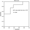
Figure 3
Receiver Operating Curve for prediction of pathologic CR rate following thermochemoradiotherapy, using change in FDG PET uptake after two weeks of treatment (29). The receiver operating curve assesses the true positive rate (Y axis) as a function of the false positive rate (X-axis). An ideal test would yield a true positive rate of 1.0, and a false positive rate of 0. Data reproduced with permission from the author and publisher.
Figure 4
Figure 4
(A). Probability of metastasis free survival as a function of time for dogs with tumors having extracellular pH>7 vs dogs with tumors having extracellular pH<7 (37). Dogs with tumors having a more alkaline extracellular pH had longer metastasis free survival.
Figure 5
Figure 5
Metastasis free survival in dogs with tumors dichotomized by contrast medium washin rate (A) and contrast medium washout rate (B), as determined from DCE-MRI (46). Metastasis free survival was longer in dogs with higher washin and washout values, likely reflecting tumors with better perfusion. The Kaplan Meier curves shown compare the survival for animals, grouped above and below the median for the whole population. Washin value is a rate constant derived from the initial slope of the dynamic contrast enhanced image set and is influenced most strongly by the perfusion rate in the tumor (10). The washout value is derived from the terminal slope of the image set and reflects the rate of contrast clearance from the tumor. This is also influenced by perfusion, but is also reflective of the extracellular volume and permeability of the microvasculature.
Figure 6
Figure 6
Hemoglobin saturation is plotted as a function of time after treatment with MTD doxorubicin. The bars represent the mean and standard deviations of the treated (blue) and control (red) groups. The dashed lines show linear regression lines for each group. A significant difference in the longitudinal trend associated with treatment was found using a linear mixed effects model (p<0.001).

Similar articles

Cited by

References

    1. Garman KS, Nevins JR, Potti A. Genomic strategies for personalized cancer therapy. Hum Mol Genet. 2007 Oct 15;16 Spec No. 2:R226–32. - PubMed
    1. Swanton C, Szallasi Z, Brenton JD, Downward J. Functional genomic analysis of drug sensitivity pathways to guide adjuvant strategies in breast cancer. Breast Cancer Res. 2008;10(5):214. - PMC - PubMed
    1. Bonnefoi H, Potti A, Delorenzi M, Mauriac L, Campone M, Tubiana-Hulin M, et al. Validation of gene signatures that predict the response of breast cancer to neoadjuvant chemotherapy: a substudy of the EORTC 10994/BIG 00-01 clinical trial. Lancet Oncol. 2007 Dec;8(12):1071–8. - PubMed
    1. Goldstein LJ, Gray R, Badve S, Childs BH, Yoshizawa C, Rowley S, et al. Prognostic utility of the 21-gene assay in hormone receptor-positive operable breast cancer compared with classical clinicopathologic features. J Clin Oncol. 2008 Sep 1;26(25):4063–71. - PMC - PubMed
    1. Mendiratta P, Febbo PG. Genomic signatures associated with the development, progression, and outcome of prostate cancer. Mol Diagn Ther. 2007;11(6):345–54. - PubMed

LinkOut - more resources