. 2010 Apr 1;201(7):1084-95.
doi: 10.1086/651172.

BBA52 facilitates Borrelia burgdorferi transmission from feeding ticks to murine hosts

Affiliations

BBA52 facilitates Borrelia burgdorferi transmission from feeding ticks to murine hosts

Manish Kumar et al. J Infect Dis. .

Abstract

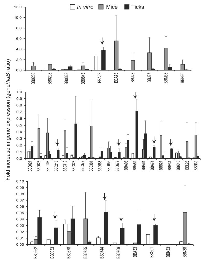
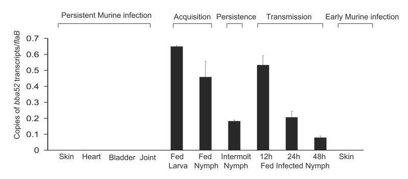
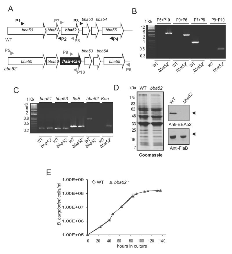
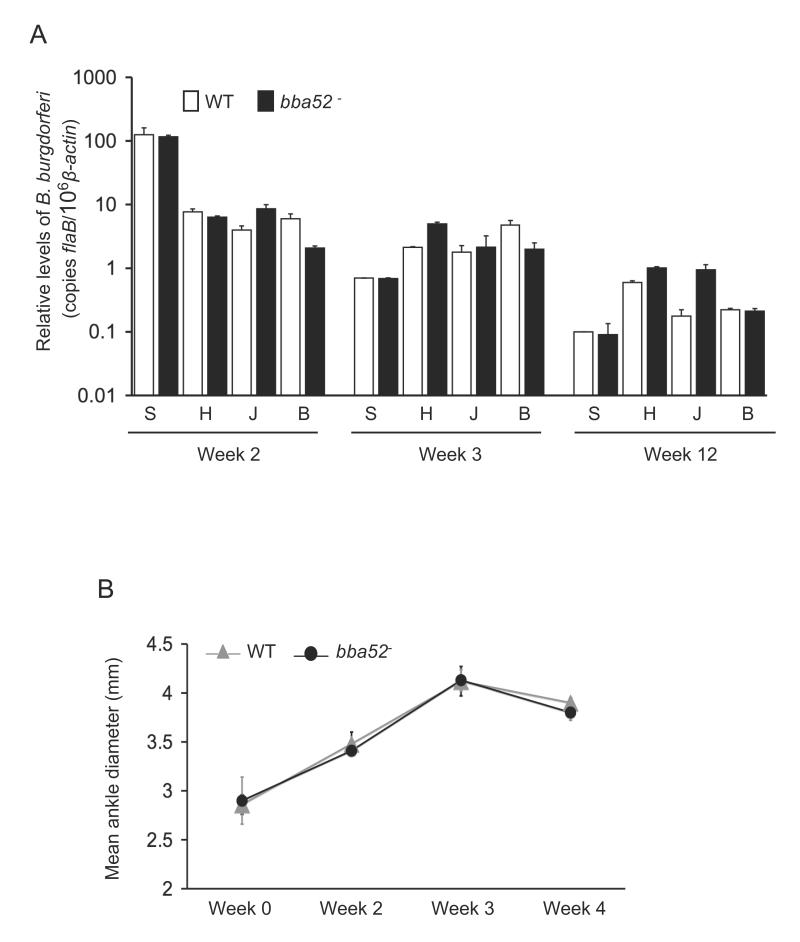
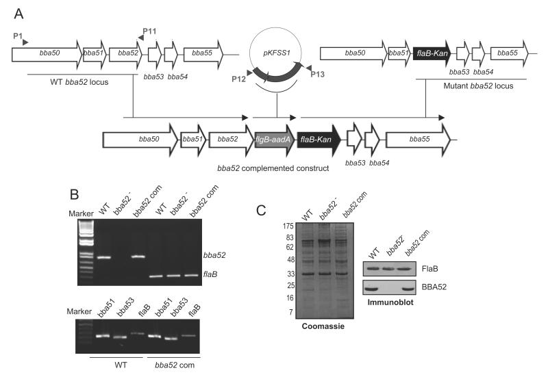
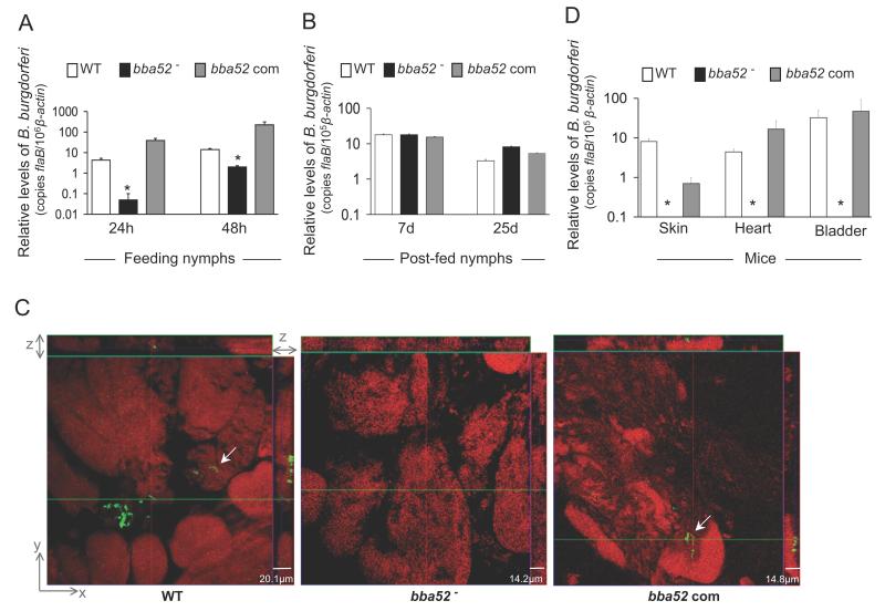
Borrelia burgdorferi, the pathogen of Lyme borreliosis, persists in nature through a tick-rodent transmission cycle. A selective assessment of the microbial transcriptome, limited to gene-encoding putative membrane proteins, reveals that bba52 transcription in vivo is strictly confined to the vector-specific portion of the microbial life cycle, with the highest levels of expression noted in feeding ticks and with swift down-regulation noted in mice. bba52 deletion did not affect murine disease as assessed by the genesis of arthritis and carditis or long-term persistence of pathogens in mice or ticks. However, bba52 deficiency did impair microbial transitions between hosts and vector, defects that could be fully rescued when bba52 expression was genetically restored to the original genomic locus. These studies establish that BBA52 facilitates vector-host transitions by the pathogen and therefore is a potential antigenic target for interference with transmission of B. burgdorferi from ticks to mammalian hosts.


Figures

Figure 1
Figure 1. Relative expression levels of selected B. burgdorferi genes in feeding ticks during transmission
Total RNA was isolated from cultured spirochetes, from pooled B. burgdorferi infected nymphs collected at 24, 48 and 96 hours of feeding on naïve mice and from murine skin following 2 weeks of B. burgdorferi infection, and converted to cDNA for measuring gene-specific transcripts using qRT-PCR. Fold increase in the expression of individual genes in each of the tick or murine samples was calculated based on threshold cycle (Ct) values using the 2−ΔΔCt method normalized against flaB Ct values. Upper, middle and lower panels represent genes with the highest, moderate and lowest expression ratios relative to that of flaB. Bars indicate the mean ± SD from four qRT-PCR analyses of two independent infection experiments. Arrows indicates genes that are highly expressed in feeding ticks but remain undetectable in the murine dermis.
Figure 2
Figure 2. bba52 expression is vector-specific
bba52 expression is analyzed at various stages of murine and tick infectivity. RNA was isolated from mice (10 animals/group) at 1, 2 and 3 weeks after B. burgdorferi infection and pooled by the tissue types (skin, heart, joint and bladder). Naïve larvae and nymphs were allowed to feed on B. burgdorferi-infected mice (25 ticks/mice) and collected at 96 hours (fed larva and fed nymph) or 25 days following feeding (intermolt nymph). B. burgdorferi-infected nymphs (10 nymphs/mouse) were allowed to feed on naïve mice and collected at 12, 24 and 48 hours of feeding (fed infected nymph). Murine skin samples were collected following 5 days of tick engorgement. RNA samples from murine and tick samples were analyzed by qRT-PCR and presented as copies of bba52 transcript per copy of flaB transcript. Error bars represent the mean ± SEM from four qRT-PCR analyses of two independent murine-tick infection experiments.
Figure 3
Figure 3. Construction and analysis of bba52 mutant B. burgdorferi
(A) Schematic representation of wild type (WT) and bba52 mutant (bba52-) B. burgdorferi at the bba52 locus. Genes bba50-bba55 (white box arrows) and the kanamycin-resistance cassette driven by the B. burgdorferi flaB promoter (flaB-Kan, black box arrow) are indicated. The regions up- and down-stream of the bba52 locus were amplified using primers P1-P4 (black arrow-heads) and ligated on either side of the flaB-Kan cassette to obtain the mutagenic construct, as detailed in the text. (B) Integration of the mutagenic construct, flaB-Kan, in the intended genomic locus. Primers 5-10 (gray arrowheads, positions indicated in figure 3A) were used for PCR analysis using isolated DNA from wild type (WT) or mutant B. burgdorferi (bba52-) and subjected to gel electrophoresis. The combination of primers used for PCR is indicated at the top, and migration of the DNA ladder is shown on the left. (C) RT-PCR assessment of bba52 transcripts and the polar effects of mutagenesis. Total RNA was isolated from wild-type B. burgdorferi (WT) and bba52 mutant (bba52-), converted to cDNA and used to amplify regions within bba52, flaB, kanamycin and genes surrounding the bba52 locus (bba51 and bba53) and visualized on a gel. (D) Protein analysis of wild-type B. burgdorferi (WT) and bba52 mutant (bba52-). Equal amounts of protein were separated on an SDS-PAGE gel, and either stained with Coomassie blue (left panel) or transferred onto a nitrocellulose membrane and probed with BBA52 and FlaB antibodies (right panels). Migration of protein standards is shown to the left in kDa. (E) Growth curves for the wild-type and bba52 mutant B. burgdorferi. Spirochetes were diluted to a density of 105 cells/ml and grown at 34°C in BSK-H medium. Triplicate samples were counted under a dark-field microscope using a Petroff-Hausser cell counter. Differences between wild type and bba52 mutant numbers were insignificant at all times of growth (P > 0.05).
Figure 4
Figure 4. bba52 mutant B. burgdorferi retain full infectivity in mice
(A) The pathogen burdens in multiple tissues of infected mice are shown. Mice (15 animals/group) were infected with wild type or the bba52 mutant isolates and spirochete burdens were analyzed in skin (S), heart (H), joint (J) and bladder (B) samples by measuring copies of B. burgdorferi flaB RNA at 2, 3 and 12 weeks of infection. Amounts of murine β-actin were determined in each sample and used to normalize the quantities of spirochete RNA. Bars represent the mean measurements ± SEM of qRT-PCR analyses from two independent infection experiments. The difference between wild type and bba52 mutant levels was statistically insignificant at all time points and tissues (P > 0.05). (B) Assessment of joint swelling in B. burgdorferi-infected mice. Groups of mice (3 animals/group) were infected with wild type or bba52 mutant and examined for joint swelling, using a digital caliper at 0, 2, 3 and 4 weeks after spirochete challenge. Data represent the mean ± SEM from two independent infection experiments. No difference in the ability of the wild type and bba52 mutant to induce joint swelling was recorded (P > 0.05).
Figure 5
Figure 5. Genetic complementation of bba52 mutant B. burgdorferi
(A) Construction of the bba52-complemented construct for re-insertion of bba52 in cis, in the original gene locus of the lp54 plasmid. A new 5′ arm was generated using primers P1-P11 and P12-P13, which were used to amplify and assemble two DNA inserts surrounding bba52 and aadA cassette with the flgB promoter, respectively. The insert representing P1-P13 amplicon (new 5′ arm) was fused with the flaB-Kan cassette carrying the old 3′ arm (as generated using P3-P4, figure 3A) to obtain bba52 complemented construct, and integrated in B. burgdorferi lp54 locus via homologous recombination. (B) RT-PCR analysis of the bba52 complemented isolate. Total RNA was isolated from either the wild type (WT), bba52 mutant (bba52-) or bba52-complemented B. burgdorferi (bba52 Com), converted to cDNA, then subjected to PCR analysis with flaB and bba52 primers, and analyzed on a 1.5% agarose gel (upper panel). bba52 complemented isolates did not display polar effects on the transcription of genes surrounding bba52 locus (bba51 and bba53) (lower panel). (C) Production of BBA52 protein in the complemented B. burgdorferi. Spirochete lysates were separated on a SDS-PAGE gel, stained with Coomassie blue (left panel), or transferred to nitrocellulose membrane and probed with BBA52 and FlaB antibodies (right panels).
Figure 6
Figure 6. bba52 mutant B. burgdorferi is impaired in its ability to transit between murine hosts and ticks
(A) B. burgdorferi burdens in ticks during acquisition from infected mice. Mice were infected with B. burgdorferi (3 mice/group) and, following 2 weeks of infection, naïve I. scapularis larvae or nymphs (25 ticks/mouse) were allowed to feed on mice. B. burgdorferi burdens in ticks were analyzed at the indicated time intervals following feeding by measuring copies of the B. burgdorferi flaB RNA. Amounts of tick β-actin were determined in each sample and used to normalize the quantities of spirochete RNA. Bars represent the mean ± SEM of eight qRT-PCR analyses derived from two independent infection experiments. Differences in the spirochete burdens in ticks infected with bba52 mutant and those with the bba52-complemented isolates or wild-type spirochetes were significant both at 24 and (*P < 0.002) 48 hours (*P < 0.02). (B) B. burgdorferi burdens in post-fed ticks. Nymphs were allowed to engorge on infected mice as described in figure 6A and B. burgdorferi burdens in post-fed ticks were analyzed at the indicated time intervals by measuring copies of the B. burgdorferi flaB RNA and normalize against tick β-actin RNA. Bars represent the mean ± SEM of eight qRT-PCR analyses derived from two independent infection experiments. Similar burdens of bba52 mutants, wild type and bba52-complemented isolates were evident at day 7 or day 25 (P > 0.05). (C) B. burgdorferi localization in infected salivary glands during transmission. A representative image showing confocal orthogonal display of infected salivary glands in the XZ and YZ axis revealing the distribution of spirochetes through the full thickness of the 60-hour fed salivary glands is shown. The spirochetes (arrow) were labeled with FITC-labeled goat anti-B. burgdorferi antibody (shown in green) and gland morphology were revealed by labeling of acinar actin filaments using Texas Red-phalloidin (shown in red). While wild type and complemented B. burgdorferi (bba52 Com) were occasionally observed within the gland, bba52 mutant (bba52-) was consistently undetected. (D) B. burgdorferi transmission from infected ticks to mice. B. burgdorferi-infected nymphs were generated by feeding larvae on mice infected with wild type and genetically-manipulated spirochetes, as described in the text. Newly-molted B. burgdorferi-infected nymphs were allowed to feed on naïve mice (1 tick/mouse, 3 animals/group). B. burgdorferi burdens were assessed in the indicated murine tissues after one week of tick feeding by measuring copies of the B. burgdorferi flaB RNA and normalized against mouse β-actin levels. Bars represent the mean ± SEM of four qRT-PCR analyses derived from two independent animal infection experiments. * bba52 mutants were undetectable.

Similar articles

Cited by

References

    1. Piesman J, Eisen L. Prevention of Tick-Borne Diseases. Annu Rev Entomol. 2008;53:323–343. - PubMed
    1. Steere AC, Coburn J, Glickstein L. The emergence of Lyme disease. J Clin Invest. 2004;113:1093–101. - PMC - PubMed
    1. Barthold SW, DeSouza M, Fikrig E, Persing DH. Lyme borreliosis in the laboratory mouse. In: Schuster SE, editor. Lyme disease. Cold Spring Harbor Laboratory; Cold Spring Harbor, NY: 1992. pp. 223–242.
    1. Casjens S, Palmer N, van Vugt R, et al. A bacterial genome in flux: the twelve linear and nine circular extrachromosomal DNAs in an infectious isolate of the Lyme disease spirochete Borrelia burgdorferi. Mol Microbiol. 2000;35:490–516. - PubMed
    1. Fraser CM, Casjens S, Huang WM, et al. Genomic sequence of a Lyme disease spirochaete, Borrelia burgdorferi. Nature. 1997;390:580–6. - PubMed

Publication types

MeSH terms

Substances