. 2010 Feb 19:7:13.
doi: 10.1186/1476-9255-7-13.

Anti-inflammatory activity of nanocrystalline silver-derived solutions in porcine contact dermatitis

Affiliations

Anti-inflammatory activity of nanocrystalline silver-derived solutions in porcine contact dermatitis

Patricia L Nadworny et al. J Inflamm (Lond). .

Abstract

Background: Nanocrystalline silver dressings have anti-inflammatory activity, unlike solutions containing Ag+ only, which may be due to dissolution of multiple silver species. These dressings can only be used to treat surfaces. Thus, silver-containing solutions with nanocrystalline silver properties could be valuable for treating hard-to-dress surfaces and inflammatory conditions of the lungs and bowels. This study tested nanocrystalline silver-derived solutions for anti-inflammatory activity.

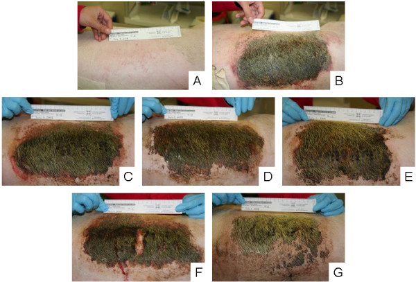
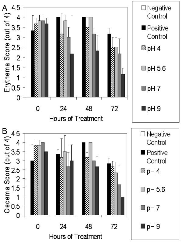
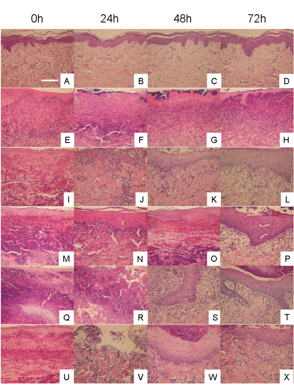
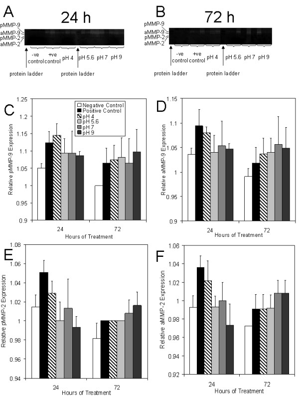
Methods: Inflammation was induced on porcine backs using dinitrochlorobenzene. Negative and positive controls were treated with distilled water. Experimental groups were treated with solutions generated by dissolving nanocrystalline silver in distilled water adjusted to starting pHs of 4 (using CO2), 5.6 (as is), 7, and 9 (using Ca(OH)2). Solution samples were analyzed for total silver. Daily imaging, biopsying, erythema and oedema scoring, and treatments were performed for three days. Biopsies were processed for histology, immunohistochemistry (for IL-4, IL-8, IL-10, TNF-alpha, EGF, KGF, KGF-2, and apoptotic cells), and zymography (MMP-2 and -9). One-way ANOVAs with Tukey-Kramer post tests were used for statistical analyses.

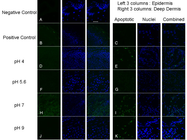
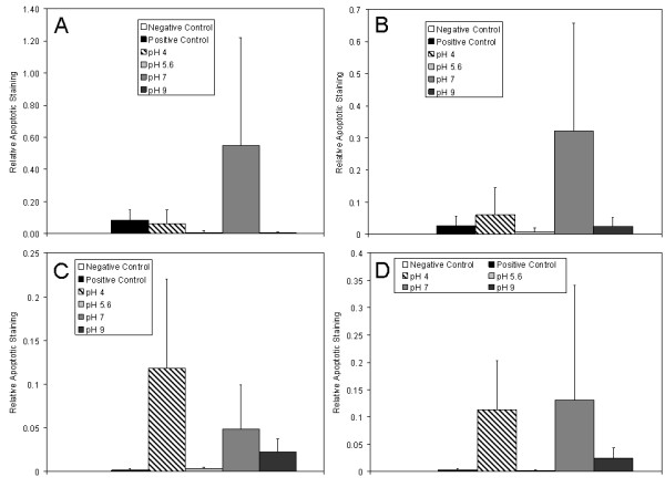
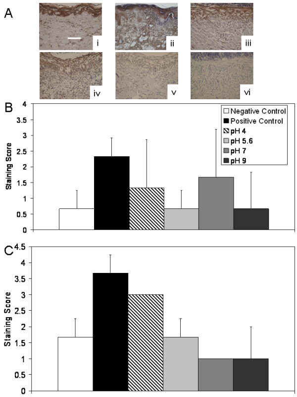
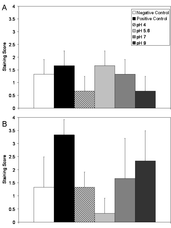
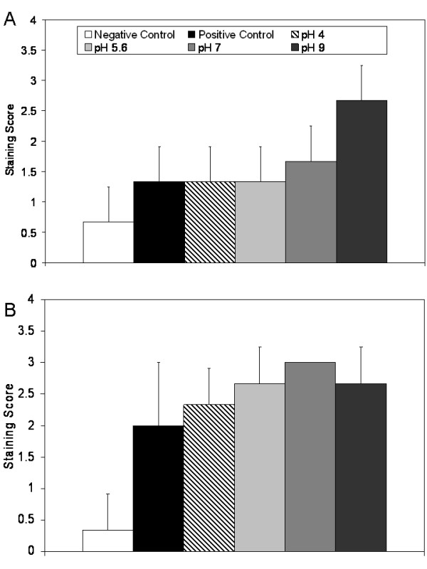
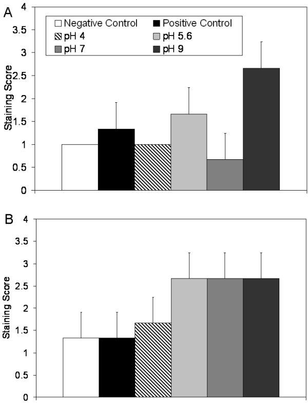
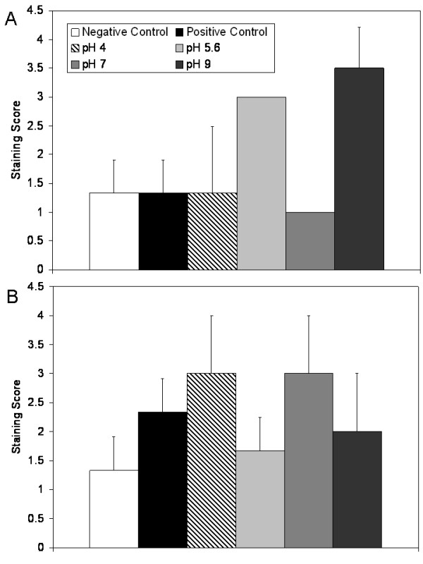
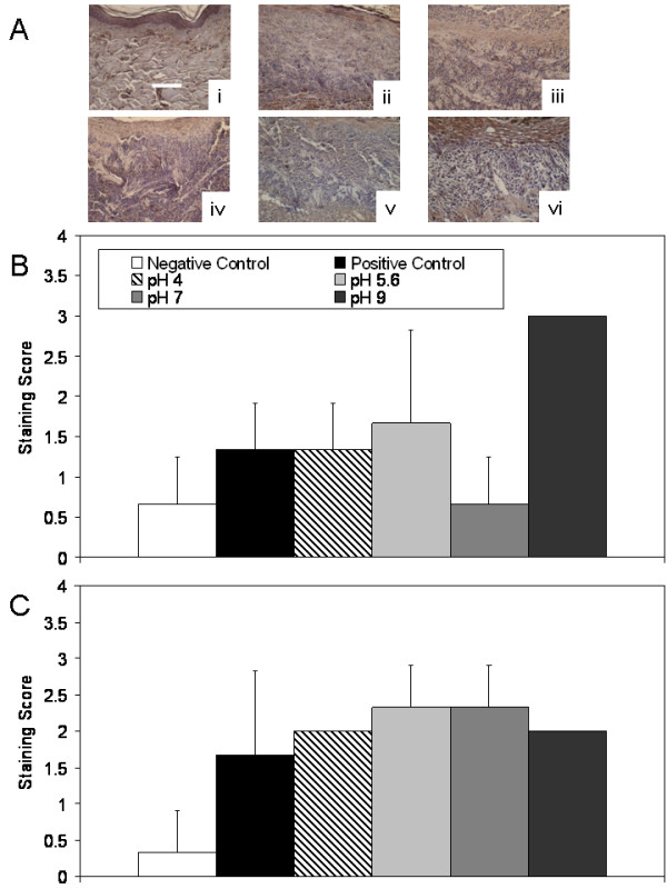
Results: Animals treated with pH 7 and 9 solutions showed clear visual improvements. pH 9 solutions resulted in the most significant reductions in erythema and oedema scores. pH 4 and 7 solutions also reduced oedema scores. Histologically, all treatment groups demonstrated enhanced re-epithelialisation, with decreased inflammation. At 24 h, pMMP-2 expression was significantly lowered with pH 5.6 and 9 treatments, as was aMMP-2 expression with pH 9 treatments. In general, treatment with silver-containing solutions resulted in decreased TNF-alpha and IL-8 expression, with increased IL-4, EGF, KGF, and KGF-2 expression. At 24 h, apoptotic cells were detected mostly in the dermis with pH 4 and 9 treatments, nowhere with pH 5.6, and in both the epidermis and dermis with pH 7. Solution anti-inflammatory activity did not correlate with total silver content, as pH 4 solutions contained significantly more silver than all others.

Conclusions: Nanocrystalline silver-derived solutions appear to have anti-inflammatory/pro-healing activity, particularly with a starting pH of 9. Solutions generated differently may have varying concentrations of different silver species, only some of which are anti-inflammatory. Nanocrystalline silver-derived solutions show promise for a variety of anti-inflammatory treatment applications.


Figures

Figure 1
Figure 1
Digital images of DNCB-induced rashes treated with various nanocrystalline silver-derived solutions. Representative digital images are shown for (A) negative controls (pigs which received no rash, and were treated with distilled water-soaked dressings); (B) DNCB-induced rashes on Day 0 before treatment was commenced; (C) positive controls (pigs which had DNCB-induced rashes and were treated with distilled water) after 72 hours of treatment; and animals treated for 72 hours with nanocrystalline silver-derived solutions with starting pHs of (D) 4, (E) 5.6, (F) 7, and (G) 9.
Figure 2
Figure 2
Erythema and oedema scores for DNCB-induced rashes treated with various nanocrystalline silver-derived solutions. Daily average erythema and oedema scores are shown in Panels A and B, respectively, for negative controls (pigs without rashes treated with distilled water-soaked dressings), and for pigs with DNCB-induced contact dermatitis treated for three days with distilled water (positive controls) or nanocrystalline silver-derived solutions with starting pHs of 4, 5.6, 7, or 9. The statistical analyses, which were performed using one-way ANOVAs with Tukey-Kramer Multiple Comparisons post tests, are shown in Table 1. Error bars represent standard deviations.
Figure 3
Figure 3
Representative histological images for DNCB-induced rashes treated with various nanocrystalline silver-derived solutions. Representative images, including portions of both the epidermis and the dermis, are shown at 0, 24, 48, and 72 h for negative controls (pigs which did not have rashes and were treated with distilled water-soaked gauze) (A-D), positive controls (pigs which had DNCB-induced rashes which were treated with distilled water-soaked gauze) (E-H), and animals with DNCB-induced rashes treated with nanocrystalline silver-derived solutions generated at starting pHs of 4 (I-L), 5.6 (M-P), 7 (Q-T), or 9 (U-X). Cell nuclei were stained purple with haematoxylin, while cytoplasm was stained pink with eosin. The scale bar in A represents 100 μm.
Figure 4
Figure 4
Gelatinase activity in biopsies from DNCB-induced rashes treated with various nanocrystalline silver-derived solutions. Zymograms are shown for all three animals of each treatment group at 24 (A) and 72 (B) hours in the following order for each time period: negative controls (no rash, treated with distilled water), positive controls (had DNCB-induced rash, treated with distilled water), and animals with DNCB-induced rashes that were treated with nanocrystalline silver-derived solutions generated at starting pHs of 4, 5.6, 7, and 9. Protein ladders were run as the first sample on each gel. The gels testing biopsies from 24 hours were run simultaneously, as were the gels testing 72 hour biopsies. The integrated density values (IDV) relative to the gel background IDV for pMMP-9, aMMP-9, pMMP-2, and aMMP-2 are shown in Panels C, D, E, and F, respectively. The statistical analyses, which were performed using one-way ANOVAs with Tukey-Kramer Multiple Comparisons Post Tests, are shown in Table 2. Error bars represent standard deviations.
Figure 5
Figure 5
Apoptosis detection in biopsies of DNCB-induced rashes treated for 24 h with various nanocrystalline silver-derived solutions. Representative fluorescence images obtained via confocal microscopy for immunohistochemical detection of apoptotic cells in pigs with DNCB-induced rashes after 24 h of various treatments are shown. The first column shows staining by FITC for apoptotic cells (green). The second column shows counterstaining by DAPI for nuclei (blue). The third column shows the combination of apoptotic and nuclear staining. Images in Row A are from the surface (epidermis and upper dermis) of a negative control (no rash) treated with distilled water. Images in Rows B and C are of the skin surface and deep dermis, respectively, of a positive control (rash treated with distilled water). Images in Rows D and E are of the surface and the deep dermis, respectively, of a DNCB-induced porcine rash treated with a nanocrystalline silver-derived solution with a starting pH of 4. Images in Rows F and G are of the surface and the deep dermis, respectively, of a DNCB-induced porcine rash treated with a nanocrystalline silver-derived solution with a starting pH of 5.6. Images in Rows H and I are of the surface and the deep dermis, respectively, of a DNCB-induced porcine rash treated with a nanocrystalline silver-derived solution with a starting pH of 7. Images in Rows J and K are of the surface and the deep dermis, respectively, of a DNCB-induced porcine rash treated with a nanocrystalline silver-derived solution with a starting pH of 9. The scale bar in the far right image in Row A represents 20 μm.
Figure 6
Figure 6
Semi-quantitative analysis of apoptosis in DNCB-induced rashes treated for 24 h with nanocrystalline silver-derived solutions. Semi-quantitative analysis of apoptotic staining in biopsies from pigs with DNCB-induced rashes after 24 h of various treatments is shown. The relative apoptotic staining level was calculated by taking a ratio of apoptotic staining (where colocalized with nuclear staining) to total nuclear staining in a given image window. Semi-quantitative analysis of staining in the epidermis, superficial dermis, deep dermis, and all dermal images combined are shown in (A) through (D), respectively. Statistical analysis, which was performed using Kruskal Wallis testing with Dunn's Multiple Comparisons post testing, is provided in Table 3. Error bars represent standard deviations.
Figure 7
Figure 7
Immunohistochemical detection of TNF-α in biopsies of DNCB-induced rashes treated with various nanocrystalline silver-derived solutions. (A) Representative images are shown for immunohistochemical detection of TNF-α after 72 h treatment of negative controls with distilled water (i), and DNCB-induced porcine contact dermatitis rashes with distilled water (positive controls) (ii), or nanocrystalline silver-derived solutions generated at starting pHs of 4 (iii), 5.6 (iv), 7 (v), or 9 (vi). The scale bar in A represents 100 μm. Staining for TNF-α appears brown, while the cell nuclei are counterstained purple using haematoxylin. Immunohistochemical staining scores for TNF-α are shown after 24 h (B) and 72 h (C) of treatment as described above. Statistical analyses, which were performed using one-way ANOVAs with Tukey-Kramer Multiple Comparisons post tests, are shown in Table 4. Error bars represent standard deviations.
Figure 8
Figure 8
Immunohistochemical detection of IL-8 in biopsies of DNCB-induced rashes treated with various nanocrystalline silver-derived solutions. Immunohistochemical staining scores for IL-8 are shown after 24 h (A) and 72 h (B) of treatment of negative controls with distilled water, and DNCB-induced porcine contact dermatitis rashes with distilled water (positive controls), or nanocrystalline silver-derived solutions generated at starting pHs of 4, 5.6, 7, or 9. Statistical analyses, which were performed using one-way ANOVAs with Tukey-Kramer Multiple Comparisons post tests, are shown in Table 4. Error bars represent standard deviations.
Figure 9
Figure 9
Immunohistochemical detection of IL-4 in biopsies of DNCB-induced rashes treated with various nanocrystalline silver-derived solutions. Immunohistochemical staining scores for IL-4 are shown after 24 h (A) and 72 h (B) of treatment of negative controls with distilled water, and DNCB-induced porcine contact dermatitis rashes with distilled water (positive controls), or nanocrystalline silver-derived solutions generated at starting pHs of 4, 5.6, 7, or 9. Statistical analyses, which were performed using one-way ANOVAs with Tukey-Kramer Multiple Comparisons post tests, are shown in Table 4. Error bars represent standard deviations.
Figure 10
Figure 10
Immunohistochemical detection of EGF in biopsies of DNCB-induced rashes treated with various nanocrystalline silver-derived solutions. Immunohistochemical staining scores for EGF are shown after 24 h (A) and 72 h (B) of treatment of negative controls with distilled water, and DNCB-induced porcine contact dermatitis rashes with distilled water (positive controls), or nanocrystalline silver-derived solutions generated at starting pHs of 4, 5.6, 7, or 9. Statistical analyses, which were performed using one-way ANOVAs with Tukey-Kramer Multiple Comparisons post tests, are shown in Table 4. Error bars represent standard deviations.
Figure 11
Figure 11
Immunohistochemical detection of KGF in biopsies of DNCB-induced rashes treated with various nanocrystalline silver-derived solutions. Immunohistochemical staining scores for KGF are shown after 24 h (A) and 72 h (B) of treatment of negative controls with distilled water, and DNCB-induced porcine contact dermatitis rashes with distilled water (positive controls), or nanocrystalline silver-derived solutions generated at starting pHs of 4, 5.6, 7, or 9. Statistical analyses, which were performed using one-way ANOVAs with Tukey-Kramer Multiple Comparisons post tests, are shown in Table 4. Error bars represent standard deviations.
Figure 12
Figure 12
Immunohistochemical detection of KGF-2 in biopsies of DNCB-induced rashes treated with various nanocrystalline silver-derived solutions. (A) Representative images are shown for immunohistochemical detection of KGF-2 after 24 h treatment of negative controls with distilled water (i), and DNCB-induced porcine contact dermatitis rashes with distilled water (positive controls) (ii), or nanocrystalline silver-derived solutions generated at starting pHs of 4 (iii), 5.6 (iv), 7 (v), or 9 (vi). The scale bar in A represents 100 μm. Staining for KGF-2 appears brown, while the cell nuclei are counterstained purple using haematoxylin. Immunohistochemical staining scores for KGF-2 are shown after 24 h (B) and 72 h (C) of treatment as described above. Statistical analyses, which were performed using one-way ANOVAs with Tukey-Kramer Multiple Comparisons post tests, are shown in Table 4. Error bars represent standard deviations.
Figure 13
Figure 13
Immunohistochemical detection of IL-10 in biopsies of DNCB-induced rashes treated with various nanocrystalline silver-derived solutions. Immunohistochemical staining scores for IL-10 are shown after 24 h (A) and 72 h (B) of treatment of negative controls with distilled water, and DNCB-induced porcine contact dermatitis rashes with distilled water (positive controls), or nanocrystalline silver-derived solutions generated at starting pHs of 4, 5.6, 7, or 9. Statistical analyses, which were performed using one-way ANOVAs with Tukey-Kramer Multiple Comparisons post tests, are shown in Table 4. Error bars represent standard deviations.

References

    1. Bhol KC, Alroy J, Schechter PJ. Anti-inflammatory effect of topical nanocrystalline silver cream on allergic contact dermatitis in a guinea pig model. Clin Exp Dermatol. 2004;29:282–287. doi: 10.1111/j.1365-2230.2004.01515.x. - DOI - PubMed
    1. Bhol KC, Schechter PJ. Topical nanocrystalline silver cream suppresses inflammatory cytokines and induces apoptosis of inflammatory cells in a murine model of allergic contact dermatitis. Br J Dermatol. 2005;152:1235–1242. doi: 10.1111/j.1365-2133.2005.06575.x. - DOI - PubMed
    1. Wright JB, Lam K, Buret AG, Olson ME, Burrell RE. Early healing events in a porcine model of contaminated wounds: effects of nanocrystalline silver on matrix metalloproteinases, cell apoptosis, and healing. Wound Repair Regen. 2002;10:141–151. doi: 10.1046/j.1524-475X.2002.10308.x. - DOI - PubMed
    1. Demling RH, DeSanti L. The rate of re-epithelialization across meshed skin grafts is increased with exposure to silver. Burns. 2002;28:264–266. doi: 10.1016/S0305-4179(01)00119-X. - DOI - PubMed
    1. Bhol KC, Schechter PJ. Topical nanocrystalline silver cream inhibits expression of MMP-9 in animal models of allergic contact dermatitis. J Invest Dermatol. 2005;124:A117.

LinkOut - more resources