. 2010 Feb 19:10:3.
doi: 10.1186/1475-2867-10-3.

Upstream molecular signaling pathways of p27(Kip1) expression: effects of 4-hydroxytamoxifen, dexamethasone, and retinoic acids

Affiliations

Upstream molecular signaling pathways of p27(Kip1) expression: effects of 4-hydroxytamoxifen, dexamethasone, and retinoic acids

Isao Eto. Cancer Cell Int. .

Abstract

Background: p27(Kip1) is a cyclin-dependent kinase inhibitor that inhibits G1-to-S phase transition of the cell cycle. It is known that a relatively large number of nutritional and chemopreventive anti-cancer agents specifically up-regulate expression of p27 without directly affecting the expression of other G1-to-S phase cell cycle regulatory proteins including p21(Cip1Waf1). However, the upstream molecular signaling pathways of how these agents up-regulate the expression of p27 have not been well characterized. The objective of this study was to identify such pathways in human breast cancer cells in vitro using 4-hydroxytamoxifen, dexamethasone, and various retinoic acids as examples of such anti-cancer agents.

Results: Experimental evidence presented in the first half of this report was obtained by transfecting human breast cancer cells in vitro with proximal upstream region of p27 gene-luciferase reporter plasmids. 1) The evidence indicated that 4-hydroxytamoxifen, dexamethasone, and various retinoic acids up-regulated expression of p27 in both estrogen receptor-positive and negative human breast cancer cells in vitro. 2) The degree of up-regulation of p27 expression by these anti-cancer agents in human breast cancer cells in vitro linearly correlated with the degree of inhibition of methylnitrosourea (MNU)-induced rat mammary adenocarcinoma in vivo. 3) Lastly, up-regulation of the expression of p27 was likely due to the activation of translation initiation rather than transcription of p27 gene. The experimental evidence presented in the second half of this report was obtained by a combination of Western immunoblot analysis and transfection analysis. It indicated that 4-hydroxytamoxifen and dexamethasone up-regulated expression of p27 by down-regulating phosphorylation of eukaryotic translation initiation factor 4E (eIF4E)-binding protein 1 (4E-BP1) at Ser65 and this phosphorylation was likely to be mediated by upstream receptor tyrosine kinases/phosphoinositide-3-kinase/Akt/5'-AMP-activated protein kinase/mammalian target of rapamycin (RTKs/PI3K/Akt/AMPK/mTOR) protein kinase signaling pathways. Retinoic acids up-regulated expression of p27 without using either 4E-BP1 or RTKs/PI3K/Akt/AMPK/mTOR protein kinase signaling pathways.

Conclusions: 4-Hydroxytamoxifen and dexamethasone up-regulated translation initiation of p27 by down-regulating 4E-BP1 phosphorylated at Ser65 and this down-regulation seemed to be mediated by upstream RTKs/PI3K/Akt/AMPK/mTOR protein kinase signaling pathways. Retinoic acids also up-regulated translation initiation of p27, but without using any of these pathways.


Figures

Figure 1
Figure 1
4-Hydroxytamoxifen, dexamethasone, all-trans-retinoic acid and 9-cis-retinoic acid up-regulate expression of p27 in both estrogen receptor-positive and -negative human breast cancer cells in vitro. (a) Outline of how various anti-cancer agents specifically up-regulate expression of p27 and arrest the progression of cell cycle from G1 to S phase. (b) Schematic drawing of the luciferase reporter plasmid containing proximal 5'-upstream region (-1797) of the p27 gene (-1797 p27 (p27-Kpn I)). 4-Hydroxytamoxifen (but not tamoxifen), dexamethasone, all-trans-retinoic acid and 9-cis-retinoic acid up-regulated relative luciferase activity of p27 in (c) estrogen receptor (ER)-positive MCF7 and (d) estrogen receptor (ER)-negative MDA-MB-231 human breast cancer cells in vitro. (e) Western immunoblot analysis of the expression of p27 protein in estrogen receptor (ER)-negative MDA-MB-231 human breast cancer cells in vitro. In all experiments, the cells were exposed to 1 μM each of tamoxifen, 4-hydroxytamoxifen, dexamethasone, all-trans-retinoic acid or 9-cis-retinoic acid for 24 hours. All assays were performed in triplicates and repeated three times.
Figure 2
Figure 2
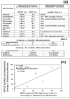
The degree of up-regulation of p27 expression in human breast cancer cells in vitro linearly correlates with the degree of inhibition of methylnitrosourea (MNU) -induced rat mammary adenocarcinoma in vivo. (a) Various chemically synthesized retinoic acids were used to investigate whether the degree of up-regulation of -1797 p27 gene-luciferase reporter (p27-Kpn I) activity in estrogen receptor (ER)-negative MDA-MB-231 human breast cancer cells in vitro correlates with the degree of inhibition of methylnitrourea (MNU) -induced rat mammary adenocarcinoma in vivo [33]. The cells were exposed to 1 μM each of the retinoic acids for 24 hours. In vitro transfection assays were performed in triplicates and repeated three times. (b) Chemical structure of the retinoic acids used in this experiment [33]. (c) Graphical representation of the results in Figure 2a above.
Figure 3
Figure 3
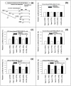
Deletion analysis indicates that 4-hydroxytamoxifen, dexamethasone, all-trans-retinoic acid (atRA) and 9-cis-retinoic acid (9cRA) activate the proximal 5'-upstream region (-1797) of p27 gene through its 5'-untranslated region (5'-UTR) (-575). (a) The 5'-deletion mutants of -1797 p27 (p27-Kip I) used in this experiment were -774 p27 (p27-Apa I) and -575 p27 (p27-5'UTR). Two additional deletion mutants, -435 p27 (p27-MB) and -417 p27 (p27-IRES), were also used in this experiments, but the data are not shown. The estrogen receptor (ER)-negative MDA-MB-231 human breast cancer cells were transfected with these deletion mutants and then exposed to 1 μM each of (b) tamoxifen, (c) 4-hydroxytamoxifen (4-OH-tamoxifen), (d) dexamethasone, (e) all-trans-retinoic acid (atRA) and (f) 9-cis-retinoic acid (9cRA) for 24 hours. All assays were performed in triplicates and the transfection experiments were repeated three times.
Figure 4
Figure 4
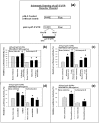
The 5'-untranslated region (5'-UTR) (-575) of p27 gene is unlikely to contain cryptic transcription factor binding sites. (a) Schematic drawing (adapted from the references [11,34]) of the pGL3-control-p27-5'-UTR-luciferase reporter plasmid. (b) The ER-negative MDA-MB 231 cells were transfected with -575 p27 (p27-5'-UTR) -luciferase reporter plasmid and then treated with either vehicle or actinomycin D(0.5 μg/ml) [55]. One hour after the addition of either vehicle or actinomycin D, the cells were exposed to vehicle, tamoxifen (1 μM) or 4-hydroxytamoxifen (1 μM) for another 24 hours. (c) Same as in Figure 4b above, except that the cells were exposed to vehicle, all-trans-retinoic acid (atRA) (1 μM) or 9-cis-retinoic acid (9cRA) (1 μM) for 24 hours. (d) Same as in Figure 4b above, except that the cells were exposed to vehicle, 4-methyl-UAB30 (4meUAB30) (1 μM) or UAB30 (1 μM) for 24 hours. (e) Same as in Figure 4b, except that the cells were exposed to vehicle or dexamethasone (1 μM). All assays were performed in triplicates and the transfection experiments were repeated three times.
Figure 5
Figure 5
4-Hydroxytamoxifen and dexamethasone up-regulate the expression of p27 by down-regulating phosphorylation of 4E-BP1 and this down-regulation is likely to be mediated by upstream Akt/AMPK/mTOR protein kinase signaling pathways. Retinoic acids also up-regulate the expression of p27 but they do so without using any of these pathways. (a) and (b): Schematic drawings of the four upstream molecular signaling pathways of p27 expression identified in our previous study [10]. These pathways are: pathway #1 (Figures 5a and 5b), pathway #2 (Figures 5a and 5b), pathway #3 (Figure 5b) and pathway #4 (Figure 5b). The specific inhibitors and activators used previously to identify these four pathways are indicated next to each of the four pathways. From (c) to (f): Estrogen receptor (ER) -negative MDA-MB-231 human breast cancer cells in vitro were exposed to vehicle, tamoxifen (1 μM), 4-hydroxytamoxifen (1 μM), dexamethasone (1 μM), all-trans-retinoic acid (atRA) (1 μM) or 9-cis-retinoic acid (9cRA) (1 μM) for 24 hours. Western immunoblot assays of the cells exposed to these anti-cancer agents were performed using antibodies against (c) total 4E-BP1, (d) 4E-BP1 phosphorylated at Ser65, (e) total AMPK, and (f) AMPK phosphorylated at Thr172. All assays were performed in triplicates and repeated three times. Abbreviations: RTK, receptor tyrosine kinase; PI3K, phosphoinositide 3-kinase; PKB, protein kinase B; AMPK, 5'-AMP-activated protein kinase; TSC, tuberous sclerosis complex; mTOR, mammalian target of rapamycin; eIF4E, eukaryotic translation initiation factor 4E; 4E-BP1, eIF4E-binding protein 1; MAPK, mitogen-activated protein kinase; Raf, MAP kinase kinase kinase; MEK, MAP kinase kinase; MKK, MAP kinase kinase; MNK, MAP kinase-interacting kinase; eIF2α, eukaryotic translation initiation factor 2α.
Figure 6
Figure 6
Further results (continuation of Figure 5) of the Western immunoblot analysis. Same as in Figure 5, except that Western immunoblot assays of the cells were performed using antibodies against (a) total Akt/PKB, (b) Akt/PKB phosphorylated at Thr308, (c) total IRS-1 (insulin receptor substrate 1), IRS-1 phosphorylated at Ser636/639, total PTEN (phosphatase and tensin homolog), PTEN phosphorylated at Ser380, total eIF4E (eukaryotic translation initiation factor 4E), eIF4E phosphorylated at Ser209, total eIF2α (eukaryotic translation initiation factor 2α), and eIF2α phosphorylated at Ser52, (d) PDGFRβ (platelet-derived growth factor receptor b) phosphorylated at Tyr751, and (e) p44/42 MAPK or ERK1/2 phosphorylated at Thr202Tyr204. All assays were performed in triplicates and repeated three times.
Figure 7
Figure 7
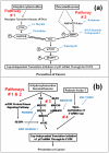
Schematic drawing of the four upstream molecular signaling pathways of p27 expression that could lead to activation of the unusually long 5'-untranslated region (5'-UTR) (-575) of p27 mRNA by 4-hydroxytamoxifen, dexamethasone and retinoic acids. (a) The two upstream molecular signaling pathways of p27 shown in Figure 5a are pathways #1 and #2. The pathway #1 consisted of receptor tyrosine kinases/phosphoinositide 3-kinase/Akt/tuberous sclerosis complex/mammalian target of rapamycin/eukaryotic translation initiation factor 4E (eIF4E) -binding protein 1 (RTKs/PI3K/Akt/TSC/mTOR/4E-BP1). The pathway #2 consisted of 5'-AMP-activated protein kinase (metabolic energy sensor or cellular fuel gauge)/tuberous sclerosis complex/mammalian target of rapamycin/eIF4E-binding protein 1 (AMPK/TSC/mTOR/4E-BP1). (b) In addition to these two pathways, two more upstream molecular signaling pathways of p27 expression were previously identified. They were pathways #3 and #4. The pathway #3 consisted of receptor tyrosine kinases/MAPKs/eIF4E (RTKs/MAPKs/eIF4E). The pathway #4 consisted of global hypomethylation of the 5'-7-methylguanosine (m7G) cap of mRNAs. The specific inhibitors and activators used previously to identify these four pathways are indicated next to each of the four pathways. The results of this study suggested that 4-hydroxytamoxifen used pathway #1 and dexamethasone primarily used pathway #2 to up-regulate the expression of p27. Dexamethasone could also use a portion of pathway #1 secondarily. We also believe, but could not conclude, that 4-hydroxytamoxifen up-regulated the expression of p27 using MAP kinase pathways (Pathway #3 in Figures 5b and 7b). Retinoic acids up-regulated p27 expression without using pathways #1, #2 and #3. We propose a hypothesis that retinoic acids are likely to have used pathway #4 to up-regulate the expression of p27. Abbreviations: see the legend of Figure 5.
Figure 8
Figure 8
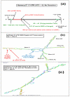
Diagrams showing the overall primary and secondary structures of the 5'-untranslated region (5'-UTR) (-575) of human p27 mRNA. (a) Overall primary structure of human 5'-UTR of p27 gene showing the location of upstream open reading frame (uORF) (-521 to -425) and the polypyrimidine tract (-66 to -41) in the internal ribosome entry site (IRES) (i.e., IRES region of Hengst: -417 to -1) [11,34]. (b) The most stable secondary stem and loop structure of the wild-type IRES region of Hengst (-417 to -1) [11,34-36] at several lowest Gibbs free energy values. This structure was generated using Zucker's RNA mfold software version 2.3 [56,57]. (c) Same as in Figure 8b, except that UU at -57 and -56 in the polypyrimidine tract (-66 to -41) were mutated to GG. This mutation completely destroyed the overall secondary stem and loop structure of IRES. Various other mutations introduced into the polypyrimidine tract also significantly modified the overall stem and loop structure of IRES.

Similar articles

Cited by

References

    1. Alkarain A, Slingerland J. Deregulation of p27 by oncogenic signaling and its prognostic significance in breast cancer. Breast Cancer Res. 2004;6:13–21. doi: 10.1186/bcr722. - DOI - PMC - PubMed
    1. Morgan DO. Principles of CDK regulation. Nature. 1995;374:131–134. doi: 10.1038/374131a0. - DOI - PubMed
    1. Sherr CJ, Roberts JM. Inhibitors of mammalian G1 cyclin-dependent kinases. Genes Dev. 1995;9:1149–1163. doi: 10.1101/gad.9.10.1149. - DOI - PubMed
    1. Sherr CJ, Roberts JM. CDK inhibitors: positive and negative regulators of G1-phase progression. Genes Dev. 1999;13:1501–1512. doi: 10.1101/gad.13.12.1501. - DOI - PubMed
    1. Slingerland J, Pagano M. Regulation of the cdk inhibitor p27 and its deregulation in cancer. J Cell Physiol. 2000;183:10–17. doi: 10.1002/(SICI)1097-4652(200004)183:1<10::AID-JCP2>3.0.CO;2-I. - DOI - PubMed