. 2010 Feb 19:10:19.
doi: 10.1186/1471-213X-10-19.

SEL1L deficiency impairs growth and differentiation of pancreatic epithelial cells

Affiliations

SEL1L deficiency impairs growth and differentiation of pancreatic epithelial cells

Shuai Li et al. BMC Dev Biol. .

Abstract

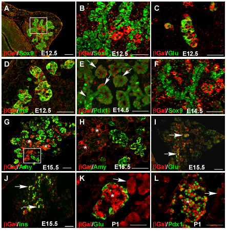
Background: The vertebrate pancreas contains islet, acinar and ductal cells. These cells derive from a transient pool of multipotent pancreatic progenitors during embryonic development. Insight into the genetic determinants regulating pancreatic organogenesis will help the development of cell-based therapies for the treatment of diabetes mellitus. Suppressor enhancer lin12/Notch 1 like (Sel1l) encodes a cytoplasmic protein that is highly expressed in the developing mouse pancreas. However, the morphological and molecular events regulated by Sel1l remain elusive.

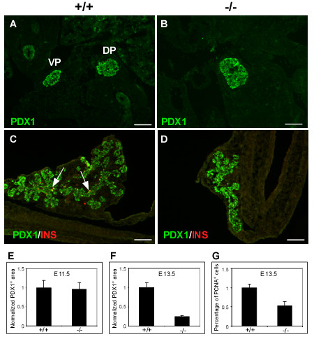
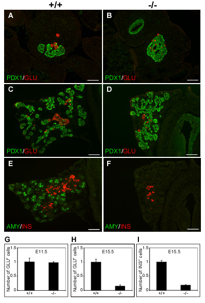
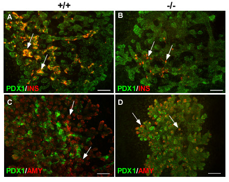
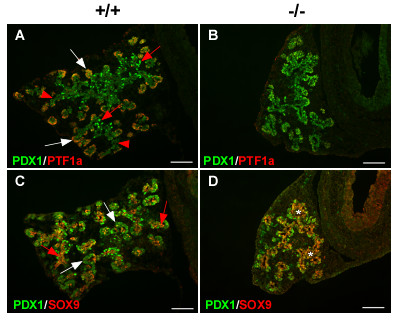
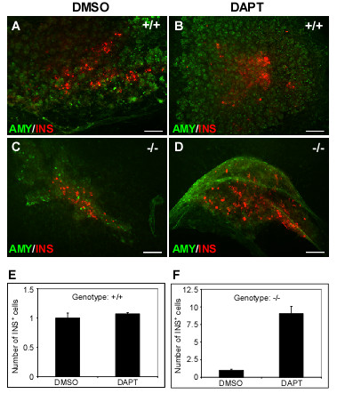
Results: We have characterized the pancreatic phenotype of mice carrying a gene trap mutation in Sel1l. We show that Sel1l expression in the developing pancreas coincides with differentiation of the endocrine and exocrine lineages. Mice homozygous for the gene trap mutation die prenatally and display an impaired pancreatic epithelial morphology and cell differentiation. The pancreatic epithelial cells of Sel1l mutant embryos are confined to the progenitor cell state throughout the secondary transition. Pharmacological inhibition of Notch signaling partially rescues the pancreatic phenotype of Sel1l mutant embryos.

Conclusions: Together, these data suggest that Sel1l is essential for the growth and differentiation of endoderm-derived pancreatic epithelial cells during mouse embryonic development.


Figures

Figure 1
Figure 1
Spatiotemporal expression of Sel1l during development of the mouse pancreas. Pancreatic sections from timed Sel1l+/βgeo embryos were co-immunostained with antibodies against βGal (red) and various pancreatic markers (green) as indicated on the left side of each panel. (B and H) Magnified views of the boxed area in A and G, respectively. (A-D) βGal expression at E12.5. βGal expression begins in a small number of cells within the core pancreatic epithelium at E12.5 (A). At this stage, there is no co-expression of βGal and SOX9 (B); however, βGal is co-expressed with either glucagon (C) or insulin (D). (E-F) βGal expression at E14.5. βGal is differentially expressed in the PDX1+ cells (E). While high βGal signal was detected in the epithelial branches (arrows), no or very low Gal signal was detected in the core "duct-like" epithelium (arrowhead). These core epithelial cells correspond to the subset of PDX+ cells that express SOX9 (F). (G-J) βGal expression at E15.5. βGal expression increases markedly in the core pancreatic epithelium (G and H, asterisks). βGal co-localizes with either glucagon (I, arrows) or insulin (J, arrows). (K-L) βGal expression at postnatal day 1. βGal is expressed in both islet and acinar tissues (K, asterisk and arrow, respectively). βGal co-localizes with PDX1 in the islet tissue (L). Scale bars: (A, G, I and J) 50 μm; (B-F, K-L) 100 μm.
Figure 2
Figure 2
Impaired pancreatic epithelial growth and branching morphogenesis in Sel1lβgeo/βgeo embryos. (A-D) Immunohistological analysis of the developing pancreas of wild-type and Sel1lβgeo/βgeo embryos at E11.5 and E13.5; the genotypes of the pancreatic sections are indicated as +/+ and -/-, respectively. The following antibodies were used: Pdx1 (A-D, green) and insulin (C-D, red). (A-B) The dorsal and ventral pancreatic buds in Sel1lβgeo/βgeo embryos are fused together. (C-D) The dorsal pancreatic bud of Sel1lβgeo/βgeo embryos exhibits a markedly reduced epithelial size and an impaired branching morphology. (E-F) Statistical analyses of estimated pancreatic epithelial sizes of wild-type and Sel1lβgeo/βgeo embryos at E11.5 (E) and E13.5 (F). Data were from three sets of wild-type and Sel1lβgeo/βgeo embryos. No significant difference in the epithelial sizes of wild-type and Sel1l-deficient pancreas at E11.5 was detected (E). At E13.5, the pancreatic epithelium of Sel1l-deficient embryos was significantly smaller (F). Scale bar: 100 μm.
Figure 3
Figure 3
Inhibited endocrine and exocrine cell differentiation in Sel1lβgeo/βgeo embryos. (A-F) Immunohistological analysis of the developing pancreas of Sel1l+/+ and Sel1lβgeo/βgeo embryos at E11.5 (A-B) and E15.5 (C-F); the genotypes of the pancreatic sections are indicated as "+/+" and "-/-", respectively. The following antibodies were used: Pdx1 (A-D, green), glucagon (A-D, red), insulin (E-F, red) and amylase (E-F, green). (A-B) At E11.5, Glu+ cells were detected in the pancreatic epithelium of wild-type and Sel1lβgeo/βgeo embryos in equal numbers. (C-D) At E15.5, the number of glucagon+ cells was reduced in Sel1lβgeo/βgeo embryos as compared to wild-type control. (E-F) No amylase+ cells were detected and the number of insulin+ cells was significantly reduced in Sel1lβgeo/βgeo embryos. (G-I) Statistical analyses of the numbers of glucagon+ and insulin+ cells in wild-type and Sel1lβgeo/βgeo embryos. Data were from three wild-type and three Sel1lβgeo/βgeo embryos. At E11.5, no significant difference in the number of glucagon+ cells in wild-type and Sel1lβgeo/βgeo embryos was detected (G). The number of glucagon+ and insulin+ cells was markedly lower in Sel1lβgeo/βgeo embryos than in wild-type control embryos (H and I, respectively). Scale bar: 100 μm.
Figure 4
Figure 4
Sel1l-deficient pancreatic epithelium exhibits impaired growth, branching morphology and differentiation ex vivo. Dorsal pancreatic buds of wild-type and Sel1lβgeo/βgeo embryos were isolated at E11.5 and cultured as described in Materials and Methods. The genotypes of the pancreatic explants are indicated as "+/+" and "-/-", respectively. Cultured pancreatic explants were immunostained using the indicated antibodies. Sel1l-deficient pancreatic epithelium grows poorly and displays impaired branching morphology (B and D), as compared to wild-type control explants (A and C). The number of insulin+ cells in mutant pancreatic epithelia were markedly reduced (B, arrows), as compared to the wild-type controls (A, arrows). Amylase expression was detected in Sel1l-deficient pancreatic epithelium (D, arrows), but the expression is significantly lower than that in wild-type epithelium (C, arrows). Scale bar: 100 μm.
Figure 5
Figure 5
Sel1l-deficient pancreatic epithelial cells fail to commit to lineage precursors during the secondary transition. (A-D) Immunohistological analysis of the developing dorsal pancreas of wild-type and Sel1lβgeo/βgeo embryos at E15, using the indicated antibodies. The genotypes of the pancreatic sections are indicated as "+/+" and "-/-", respectively. (A-B) PTF1a is expressed in the periphery of wild-type pancreatic epithelium marking the commitment of pancreatic epithelial cells into the exocrine lineage (A, white arrows); PTF1a expression is absent in the mutant pancreas (B). Up-regulation of PDX1 expression in a subset of epithelial cells in the core of wild-type pancreatic epithelium marks the commitment of pancreatic epithelial cells into the endocrine lineage (A, red arrows); whereas PDX1 is uniformly expressed in mutant pancreatic epithelial cells (B). (C-D) SOX9 is expressed in a small population of epithelial cells in wild-type pancreatic epithelium (C, red arrows); in contrast, SOX9 mostly co-localizes with PDX1 in mutant pancreatic epithelium (D, asterisks). Scale bar: 100 μm.
Figure 6
Figure 6
DAPT treatment rescues the pancreatic phenotype of Sel1lβgeo/βgeo embryos. The dorsal pancreatic bud of wild-type (+/+) and Sel1l mutant (-/-) embryos was isolated at E11.5 and cultured for 8 days in the presence of 0.1% DMSO (control) and 10 μM of DAPT. (A-D) Immunohistological analysis of DAPT-treated and non-treated pancreatic buds with the indicated antibodies. (E-F) Quantification of insulin+ cells in DMSO and DAPT-treated pancreatic explants. DAPT did not significantly change the number of insulin+ cells in wild-type pancreatic buds (A-B and E), however, it induced a marked increase of insulin+ cells in Sel1l mutant pancreatic buds (C-D and F). Scale bar: 100 μm.

Similar articles

Cited by

References

    1. Slack JM. Developmental biology of the pancreas. Development. 1995;121:1569–80. - PubMed
    1. Jorgensen MC, Ahnfelt-Ronne J, Hald J, Madsen OD, Serup P, Hecksher-Sorensen J. An illustrated review of early pancreas development in the mouse. Endocr Rev. 2007;28:685–705. doi: 10.1210/er.2007-0016. - DOI - PubMed
    1. Kim SK, MacDonald RJ. Signaling and transcriptional control of pancreatic organogenesis. Curr Opin Genet Dev. 2002;12:540–7. doi: 10.1016/S0959-437X(02)00338-6. - DOI - PubMed
    1. Gu G, Dubauskaite J, Melton DA. Direct evidence for the pancreatic lineage NGN3+ cells are islet progenitors and are distinct from duct progenitors. Development. 2002;129:2447–57. - PubMed
    1. Pictet RL, Clark WR, Williams RH, Rutter WJ. An ultrastructural analysis of the developing embryonic pancreas. Dev Biol. 1972;29:436–67. doi: 10.1016/0012-1606(72)90083-8. - DOI - PubMed

Publication types

Substances