

Transmembrane potential induced on the internal organelle by a time-varying magnetic field: a model study
- PMID: 20170538
- PMCID: PMC2836366
- DOI: 10.1186/1743-0003-7-12
Transmembrane potential induced on the internal organelle by a time-varying magnetic field: a model study
Abstract
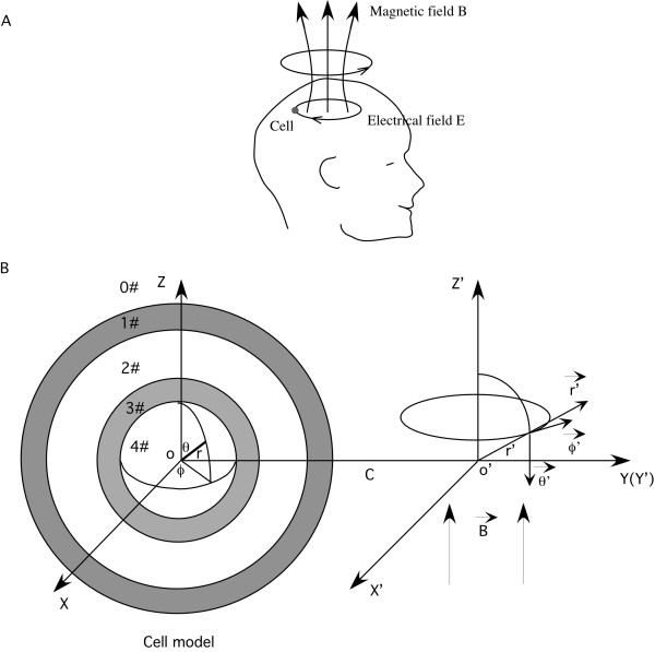
Background: When a cell is exposed to a time-varying magnetic field, this leads to an induced voltage on the cytoplasmic membrane, as well as on the membranes of the internal organelles, such as mitochondria. These potential changes in the organelles could have a significant impact on their functionality. However, a quantitative analysis on the magnetically-induced membrane potential on the internal organelles has not been performed.
Methods: Using a two-shell model, we provided the first analytical solution for the transmembrane potential in the organelle membrane induced by a time-varying magnetic field. We then analyzed factors that impact on the polarization of the organelle, including the frequency of the magnetic field, the presence of the outer cytoplasmic membrane, and electrical and geometrical parameters of the cytoplasmic membrane and the organelle membrane.
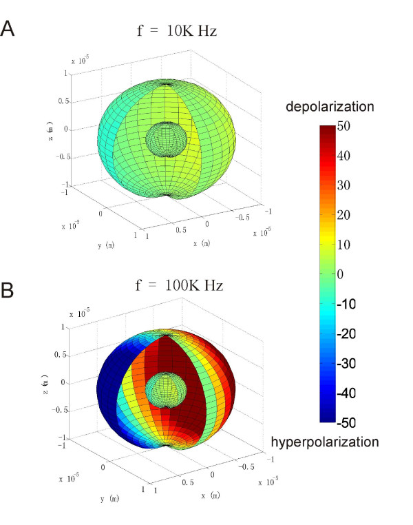
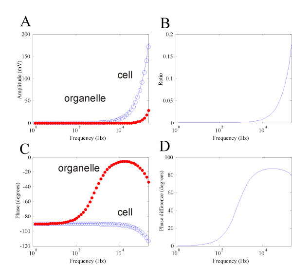
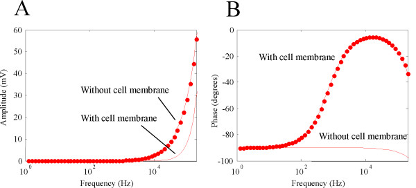
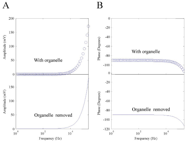
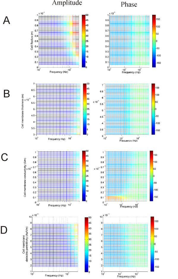
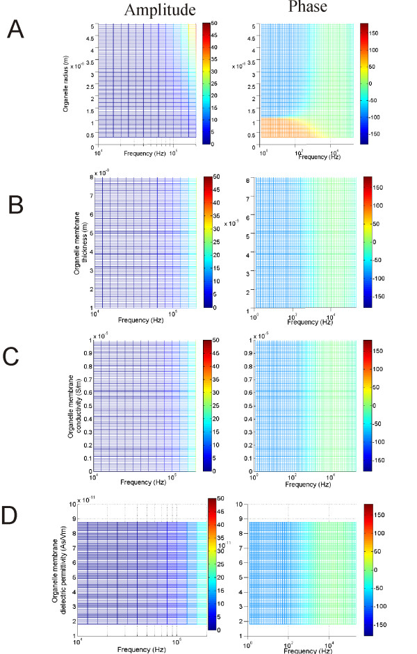
Results: The amount of polarization in the organelle was less than its counterpart in the cytoplasmic membrane. This was largely due to the presence of the cell membrane, which "shielded" the internal organelle from excessive polarization by the field. Organelle polarization was largely dependent on the frequency of the magnetic field, and its polarization was not significant under the low frequency band used for transcranial magnetic stimulation (TMS). Both the properties of the cytoplasmic and the organelle membranes affect the polarization of the internal organelle in a frequency-dependent manner.
Conclusions: The work provided a theoretical framework and insights into factors affecting mitochondrial function under time-varying magnetic stimulation, and provided evidence that TMS does not affect normal mitochondrial functionality by altering its membrane potential.
Figures
Similar articles
-
Mechanisms for the intracellular manipulation of organelles by conventional electroporation.Biophys J. 2010 Jun 2;98(11):2506-14. doi: 10.1016/j.bpj.2010.02.035. Biophys J. 2010. PMID: 20513394 Free PMC article.
-
Theoretical evaluation of voltage inducement on internal membranes of biological cells exposed to electric fields.Biophys J. 2006 Jan 15;90(2):480-91. doi: 10.1529/biophysj.105.070771. Epub 2005 Oct 20. Biophys J. 2006. PMID: 16239325 Free PMC article.
-
Transmembrane potential induced in a spherical cell model under low-frequency magnetic stimulation.J Neural Eng. 2007 Sep;4(3):283-93. doi: 10.1088/1741-2560/4/3/014. Epub 2007 Jul 3. J Neural Eng. 2007. PMID: 17873431
-
Organelle Communication at Membrane Contact Sites (MCS): From Curiosity to Center Stage in Cell Biology and Biomedical Research.Adv Exp Med Biol. 2017;997:1-12. doi: 10.1007/978-981-10-4567-7_1. Adv Exp Med Biol. 2017. PMID: 28815518 Review.
-
Power transmission along biological membranes.J Membr Biol. 1990 Mar;114(2):97-112. doi: 10.1007/BF01869092. J Membr Biol. 1990. PMID: 2111408 Review.
Cited by
-
Biomechanics of cell membrane under low-frequency time-varying magnetic field: a shell model.Med Biol Eng Comput. 2016 Dec;54(12):1871-1881. doi: 10.1007/s11517-016-1478-9. Epub 2016 Apr 6. Med Biol Eng Comput. 2016. PMID: 27053164
-
Irreversible Electroporation: Background, Theory, and Review of Recent Developments in Clinical Oncology.Bioelectricity. 2019 Dec 1;1(4):214-234. doi: 10.1089/bioe.2019.0029. Epub 2019 Dec 12. Bioelectricity. 2019. PMID: 34471825 Free PMC article. Review.
-
Restore axonal conductance in a locally demyelinated axon with electromagnetic stimulation.J Neural Eng. 2025 Feb 14;22(1):016042. doi: 10.1088/1741-2552/adb213. J Neural Eng. 2025. PMID: 39904055 Free PMC article.
-
Finding the Location of Axonal Activation by a Miniature Magnetic Coil.Front Comput Neurosci. 2022 Jun 29;16:932615. doi: 10.3389/fncom.2022.932615. eCollection 2022. Front Comput Neurosci. 2022. PMID: 35847967 Free PMC article.
-
Cellular and Molecular Effects of Magnetic Fields.Int J Mol Sci. 2024 Aug 17;25(16):8973. doi: 10.3390/ijms25168973. Int J Mol Sci. 2024. PMID: 39201657 Free PMC article. Review.
References
-
- Thompson SP. A Physiological Effect of an Alternating Magnetic Field. Proc R Soc. 1910;82:396–398. doi: 10.1098/rspb.1910.0032. - DOI
-
- Anninos PA, Tsagas N, Sandyk R, Derpapas K. Magnetic stimulation in the treatment of partial seizures. Int J Neurosci. 1991;60:141–171. - PubMed
-
- Anninos PA, Tsagas N, Jacobson JI, Kotini A. The biological effects of magnetic stimulation in epileptic patients. Panminerva Med. 1999;41:207–215. - PubMed
Publication types
MeSH terms
Grants and funding
LinkOut - more resources
Full Text Sources
Medical
