

Lifelong protection from global cerebral ischemia and reperfusion in long-lived Mclk1(+/)(-) mutants
- PMID: 20170652
- PMCID: PMC4053415
- DOI: 10.1016/j.expneurol.2010.02.002
Lifelong protection from global cerebral ischemia and reperfusion in long-lived Mclk1(+/)(-) mutants
Abstract
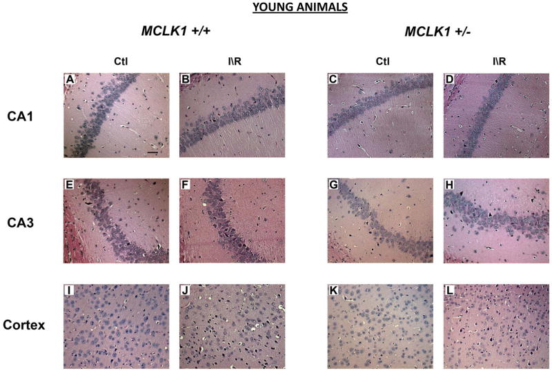
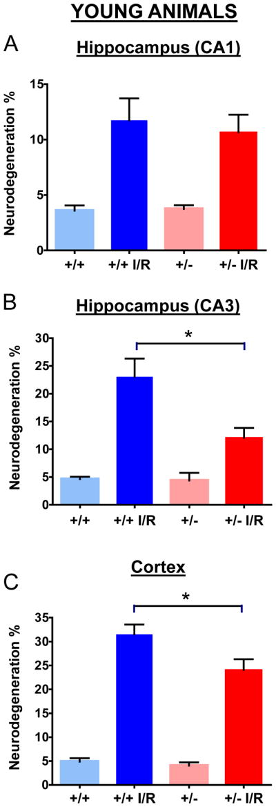
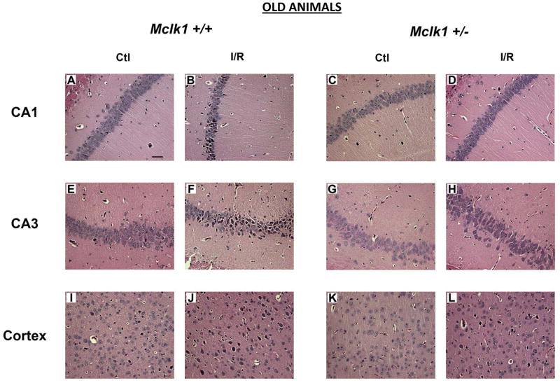
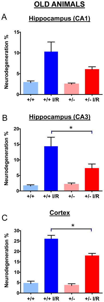
To achieve a long life span, animals must be resistant to various injuries as well as avoid or delay lethality from age-dependent diseases. Reduced expression of the mitochondrial enzyme CLK-1/MCLK1 (a.k.a. Coq7), a mitochondrial hydroxylase that is necessary for the biosynthesis of ubiquinone (UQ), extends lifespan in Caenorhabditiselegans and in mice. Here, we show that long-lived Mclk1(+/)(-) mutants have enhanced resistance to neurological damage following global cerebral ischemia-reperfusion (I/R) injury induced by transient bilateral common carotid artery occlusion (BCCAO). Both young ( approximately 100days old) and relatively aged ( approximately 450days old) mutants display increased resistance as indicated by a significant decrease in the amount of degenerating cells observed in forebrain cortex and in hippocampal areas after ischemia and reperfusion. Furthermore, less oxidative damage resulting from the procedure was measured in the brain of young Mclk1(+/)(-) animals. The finding that both young and old mutants are protected indicates that this is a basic phenotype of these mutants and not a secondary consequence of their slow rate of aging. Thus, the partial resistance to I/R injury suggests that Mclk1(+/)(-) mutants have an enhanced recovery potential following age-dependant vascular accidents, which correlates well with their longer survival. By relating this neuroprotective effect to previously reported characteristics of the Mclk1(+/)(-) phenotype, including altered mitochondrial metabolism and increased HIF-1alpha expression, this study establishes these mutants as useful models to analyze the mechanisms underlying tolerance to ischemia, particularly those associated with ischemic preconditioning, as well as to clarify the relation between aging and age-dependent diseases.
Copyright (c) 2009 Elsevier Inc. All rights reserved.
Figures
Similar articles
-
Reversal of the mitochondrial phenotype and slow development of oxidative biomarkers of aging in long-lived Mclk1+/- mice.J Biol Chem. 2009 Jul 24;284(30):20364-74. doi: 10.1074/jbc.M109.006569. Epub 2009 May 28. J Biol Chem. 2009. PMID: 19478076 Free PMC article.
-
An enhanced immune response of Mclk1⁺/⁻ mutant mice is associated with partial protection from fibrosis, cancer and the development of biomarkers of aging.PLoS One. 2012;7(11):e49606. doi: 10.1371/journal.pone.0049606. Epub 2012 Nov 14. PLoS One. 2012. PMID: 23166727 Free PMC article.
-
Early mitochondrial dysfunction in long-lived Mclk1+/- mice.J Biol Chem. 2008 Sep 19;283(38):26217-27. doi: 10.1074/jbc.M803287200. Epub 2008 Jul 17. J Biol Chem. 2008. PMID: 18635541 Free PMC article.
-
Genetic and molecular characterization of CLK-1/mCLK1, a conserved determinant of the rate of aging.Exp Gerontol. 2006 Oct;41(10):940-51. doi: 10.1016/j.exger.2006.06.041. Epub 2006 Aug 4. Exp Gerontol. 2006. PMID: 16889924 Review.
-
Brain ischemia and reperfusion: molecular mechanisms of neuronal injury.J Neurol Sci. 2000 Oct 1;179(S 1-2):1-33. doi: 10.1016/s0022-510x(00)00386-5. J Neurol Sci. 2000. PMID: 11054482 Review.
Cited by
-
The submitochondrial distribution of ubiquinone affects respiration in long-lived Mclk1+/- mice.J Cell Biol. 2012 Oct 15;199(2):215-24. doi: 10.1083/jcb.201203090. Epub 2012 Oct 8. J Cell Biol. 2012. PMID: 23045551 Free PMC article.
-
Dysregulation of iron homeostasis and methamphetamine reward behaviors in Clk1-deficient mice.Acta Pharmacol Sin. 2022 Jul;43(7):1686-1698. doi: 10.1038/s41401-021-00806-1. Epub 2021 Nov 22. Acta Pharmacol Sin. 2022. PMID: 34811513 Free PMC article.
-
Mitochondrial Retrograde Signaling: Triggers, Pathways, and Outcomes.Oxid Med Cell Longev. 2015;2015:482582. doi: 10.1155/2015/482582. Epub 2015 Oct 25. Oxid Med Cell Longev. 2015. PMID: 26583058 Free PMC article. Review.
-
Within- and between-species study of extreme longevity--comments, commonalities, and goals.J Gerontol A Biol Sci Med Sci. 2012 Apr;67(4):347-50. doi: 10.1093/gerona/gls010. Epub 2012 Mar 14. J Gerontol A Biol Sci Med Sci. 2012. PMID: 22419221 Free PMC article. No abstract available.
-
Animal Models of Coenzyme Q Deficiency: Mechanistic and Translational Learnings.Antioxidants (Basel). 2021 Oct 26;10(11):1687. doi: 10.3390/antiox10111687. Antioxidants (Basel). 2021. PMID: 34829558 Free PMC article. Review.
References
-
- Bertoni-Freddari C, Fattoretti P, Giorgetti B, Solazzi M, Balietti M, Meier-Ruge W. Role of mitochondrial deterioration in physiological and pathological brain aging. Gerontology. 2004;50:187–192. - PubMed
-
- Chan PH. Reactive oxygen radicals in signaling and damage in the ischemic brain. J Cereb Blood Flow Metab. 2001;21:2–14. - PubMed
-
- Chandel NS, Budinger GR. The cellular basis for diverse responses to oxygen. Free Radic Biol Med. 2007;42:165–174. - PubMed
-
- Cho KO, Kim SK, Cho YJ, Sung KW, Kim SY. Regional differences in the neuroprotective effect of minocycline in a mouse model of global forebrain ischemia. Life Sci. 2007;80:2030–2035. - PubMed
-
- Christophe M, Nicolas S. Mitochondria: a target for neuroprotective interventions in cerebral ischemia-reperfusion. Curr Pharm Des. 2006;12:739–757. - PubMed
Publication types
MeSH terms
Substances
Grants and funding
LinkOut - more resources
Full Text Sources
Medical
