. 2010 May;44(1):1-14.
doi: 10.1016/j.mcn.2010.01.010. Epub 2010 Feb 17.

Nervous-tissue-specific elimination of microtubule-actin crosslinking factor 1a results in multiple developmental defects in the mouse brain

Affiliations

Nervous-tissue-specific elimination of microtubule-actin crosslinking factor 1a results in multiple developmental defects in the mouse brain

Dmitry Goryunov et al. Mol Cell Neurosci. 2010 May.

Abstract

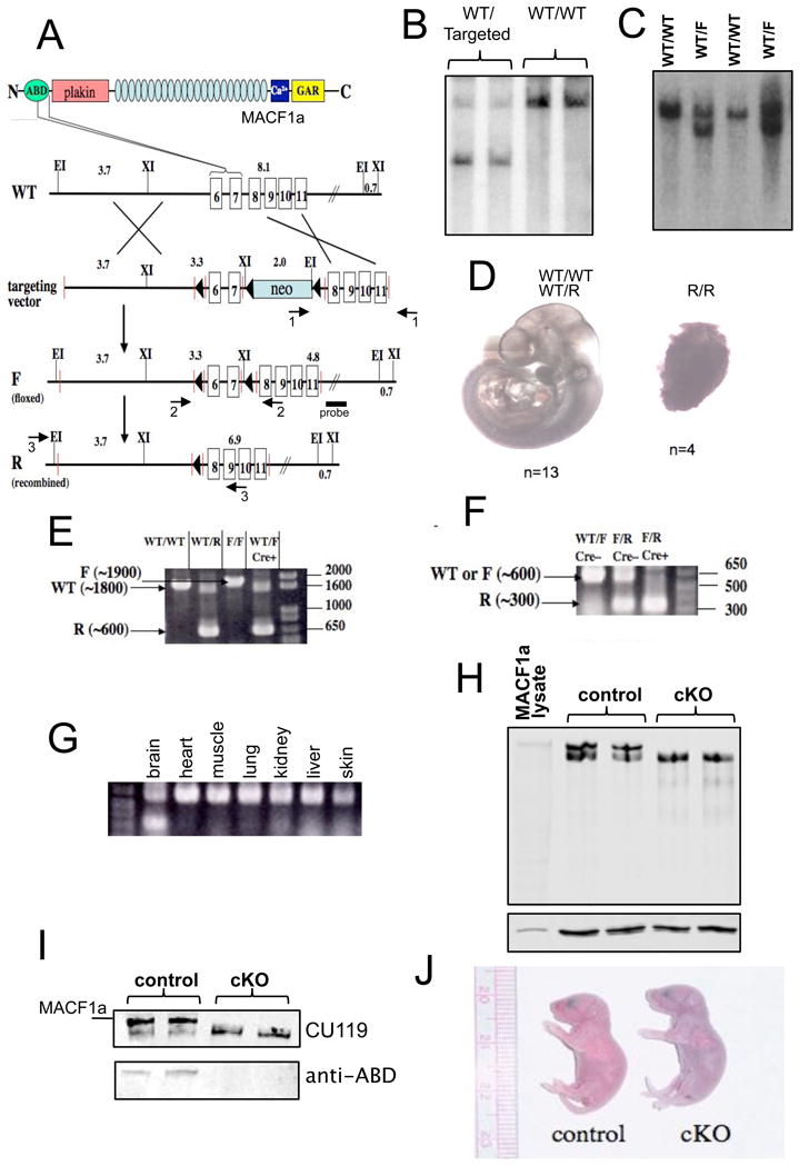
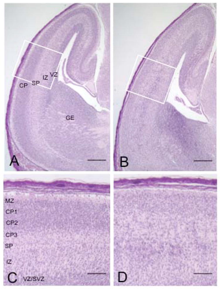
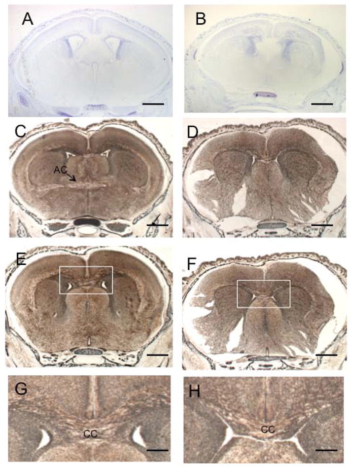
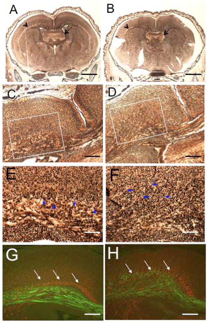
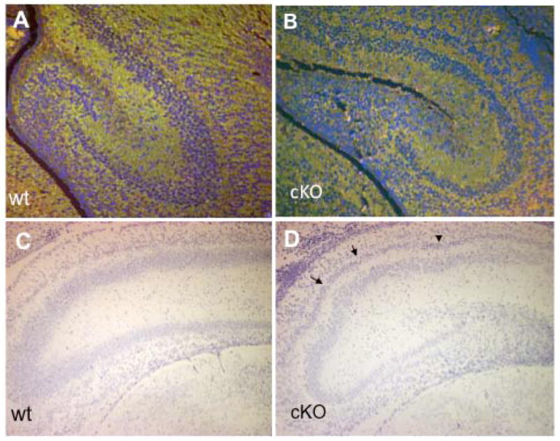
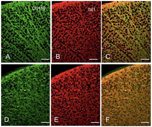
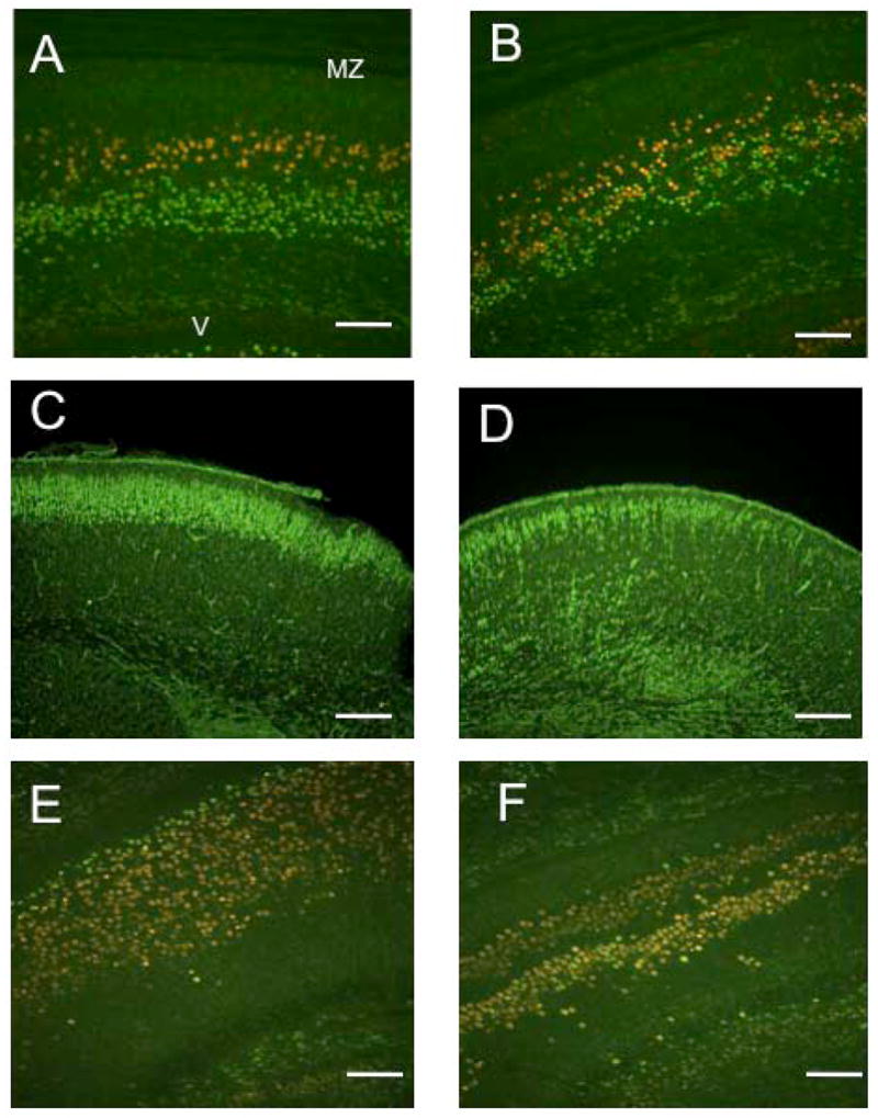
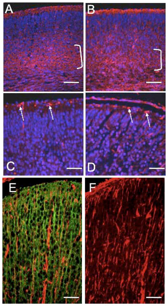
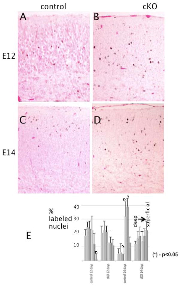
The microtubule-actin crosslinking factor 1 (MACF1) is a ubiquitous cytoskeletal linker protein with multiple spliced isoforms expressed in different tissues. The MACF1a isoform contains microtubule and actin-binding regions and is expressed at high levels in the nervous system. Macf1-/- mice are early embryonic lethal and hence the role of MACF1 in the nervous system could not be determined. We have specifically knocked out MACF1a in the developing mouse nervous system using Cre/loxP technology. Mutant mice died within 24-36h after birth of apparent respiratory distress. Their brains displayed a disorganized cerebral cortex with a mixed layer structure, heterotopia in the pyramidal layer of the hippocampus, disorganized thalamocortical and corticofugal fibers, and aplastic anterior and hippocampal commissures. Embryonic neurons showed a defect in traversing the cortical plate. Our data suggest a critical role for MACF1 in neuronal migration that is dependent on its ability to interact with both microfilaments and microtubules.


Figures

Figure 1
Figure 1
Generation of mutant Macf1 mice. (A) Schematic of the targeting strategy. Exons 6 and 7, encoding the C-terminal half of the MACF1 ABD, were floxed in the targeting construct. Recombination of this construct into the wild-type Macf1 locus resulted in a targeted Macf1 allele. The neo cassette was eliminated in vivo, resulting in a floxed allele (F). Tissue-specific Cre recombination gives rise to a fully recombined (R) allele that lacks the two exons. Primer pairs 1 and 2 were used for genotyping; pair 3, for RT-PCR. Position of the Southern blotting probe is indicated. Targeted ES cells (B) and floxed heterozygous animals (C) were genotyped by Southern blotting. Two mutant samples and 2 wild-type controls are shown in each panel. For fragment lengths see Materials and Methods. (D) Mice carrying two copies of the R allele die early in embryonic development. Two litters of E10.5 embryos resulting from Macf1+/R heterozygote matings were dissected. (E) F-to-R allele conversion in the brains of Macf1 mutant mice carrying a Cre transgene under the control of the nestin promoter was confirmed by genomic PCR. The WT, F, and R products are indicated with arrows. Note the R band, and the apparent absence of an F band, in the Macf1+/F;nes-Cre lane. RT-PCR confirmed the elimination of mRNA containing the ABD region in the brains of Macf1F/R;nes-Cre animals (F) but not in other tissues (G). (H) Western blotting of Macf1F/R (control) brain extracts with CU119 antibody revealed a high molecular weight doublet, with the top band co-migrating with plasmid-derived MACF1a (MACF1 lysate). The top band was absent in Macf1F/R;nes-Cre (cKO) brains. Lower panel shows tubulin as a loading control. (I) Re-probing the control and cKO brain extracts with a polyclonal antibody against the ABD (Antolik et al., 2007) showed that only the top band contains this domain, indicating that cKO brains no longer express MACF1a but still contain the ABD-less MACF1 protein, MACF1c. Position of plasmid-derived MACF1a is also indicated. (J) cKO mice did not have any gross abnormalities compared with control mice but died within 24–36 hrs after birth, apparently of respiratory distress. Note the cyanotic appearance of the mutant pup.
Figure 2
Figure 2
Disorganization of the cerebral cortex in Macf1 nestin-Cre animals. Sagittal sections of control (A, C) or mutant (B, D) E17.5 brains were stained with hematoxylin/eosin. The control cortex (A) contained a sharply defined cortical plate, a thin but distinct subplate, a low-cell-density intermediate zone, and a robust ventricular/subventricular zone. In contrast, the mutant cortex displayed a hypotrophic cortical plate, a reduced ventricular zone, and a loose band of cells positioned below the subplate in the normally clear intermediate zone (B). Higher magnifications also revealed a thinner marginal zone and a more even cell distribution across the cKO cortex compared with the control (C, D). CP, cortical plate; CP1–3, upper, middle, and lower tiers of the cortical plate; IZ, intermediate zone; MZ, marginal zone; SP, subplate; VZ/SVZ, ventricular/subventricular zones. Similar results were obtained from six different pairs of animals from different litters. Scale bars: A, B=250μm; C, D=50μm
Figure 3
Figure 3
Multiple developmental defects in Macf1 nestin-cKO brains. (A, B) Control (A) or mutant (B) P0 coronal sections were stained with Nissl. The anterior commissure (AC) in the cKO brain was severely aplastic. The shapes of the lateral ventricles (LV) were altered. (C–H) Control (C, E, G) or mutant (D, F, H) P0 coronal sections were silver-stained. Note the anterior commissure in the control brain (C, arrow) and its apparent absence in the cKO brain (D). The corpus callosum (CC) was greatly reduced in the cKO brain (F, H) compared to the control (E, G). G and H show higher magnifications of the white box in E and F, respectively. Similar results were obtained from three different pairs of animals from different litters. Scale bars: A–F=500μm; G,H=50μm
Figure 4
Figure 4
Thalamocortical fibers and the hippocampal commissure are abnormal in the cKO brain. (A–F) Silver-stained sections of control (A, C, E) and cKO (B, D, F) brains. The hippocampal commissure (arrows in A, B) and thalamocortical fibers (arrowheads in A, B) were significantly reduced in the cKO brains (B, D) compared to the control brains (A, C). E and F show higher magnifications of the white box in C and D, respectively. Note the much-looser structure of the cKO fibers as well as the individual axons extending tangentially into the cortical plate (E, F, arrowheads). Immunofluorescent staining of control (G) and cKO (H) sections with α-internexin and TAG-1 antibodies revealed a pronounced disorganization of the thalamocortical fibers (red) as well as corticofugal fibers (green) in the mutant brain. These results were seen with three pairs of animals from different litters. Scale bars:A, B=500μm; C, D, G, H=50μm; E, F=25μm
Figure 5
Figure 5
Pyramidal cell heterotopia in the cKO hippocampus. Control (A, C) or mutant (B, D) P0 coronal brain sections were triple-stained with CU119 antibody (green), monoclonal tubulin antibody (green), and Hoechst dye (blue; A,B), or stained with Nissl dye (C,D). Compared to the control hippocampus, the pyramidal cell layer in the mutant hippocampus was less compact and narrower (A, B). The mutant hippocampus also contained a second pyramidal layer (B, D). At a more caudal section level, the pyramidal layers in the cKO hippocampus showed undulations (D, arrowheads) that were absent in the control brain (C). Similar results were obtained from three pairs of animals from different litters. Scale bars: A–D=50μm
Figure 6
Figure 6
MACF1 expression in the mouse cortex. Coronal sections of control (A–C) or Macf1 cKO (D–F) P0 brains were stained with CU119 polyclonal antibody (A, D), monoclonal tubulin antibody (B, E), and Hoechst (C, F). C, F, superimposed double images, with MACF1 staining in green and tubulin staining in red. Similar results were obtained from three pairs of animals from different litters. Scale bars: A–F=25μm
Figure 7
Figure 7
Cortical layers are mixed in the cKO brain. Control (A) or mutant (B) coronal sections were double-stained with Ctip2 (red) and Tbr1 (green) antibodies. In the control cortices, Ctip2 and Tbr1-positive neurons formed distinct layers. In the mutant cortices, the two layers were partially mixed. Staining with Cux-1, which labels cortical layers 2–4 in the control cortex (C), shows that in the mutant cortices (D), many Cux-1 positive cells have not completed migration. Similar to the cortex, staining with Ctip2 (red) and Tbr1 (green) antibodies of control (E) and mutant (F) hippocampal sections shows a mixing of the two layer markers and also confirms the heterotopia observed before. Similar results were obtained for three pairs of animals from different litters. Scale bars: A, B, E, F=50μm; C, D=100μm
Figure 8
Figure 8
Cortical plate splitting and the radial glia are unaffected in Macf1 cKO brains. Coronal sections of control (A, C, E) or mutant (B, D, F) E17.5 brains were stained with CS-56 (A, B), reelin (C, D), or vimentin (E, F) antibodies (red), and co-stained with Hoechst nuclear dye (blue; A–D only) or CU119 antibody (green, E only). Brackets in A and B indicate subplate staining. Arrows in C and D indicate reelin-positive Cajal-Retzius cells in the marginal zone. E and F show vimentin-positive processes of radial glial cells, which did not colocalize with CU119-positive neuronal processes (E). Similar results were obtained for three pairs of animals from different litters. Scale bars: A–F=50μm
Figure 9
Figure 9
Cortical neuronal migration is delayed in Macf1 nestin-cKO brains. Pregnant Macf1F/F females were injected with BrdU at E12 (A, B) or E14 (C, D). The embryos were dissected and their brains fixed at E18. Coronal sections of control (A, C) or mutant (B, D) littermates were stained with BrdU monoclonal antibody and secondary HRP conjugates, and counterstained with eosin. Early-born neurons (E12) migrate into the deeper layers of the cortical plate in both control and mutant cortices (dark dots in A, B). In contrast, late-born neurons (E14) reach the upper levels only in the control cortex (C), whereas in the mutant cortex they are scattered throughout the cortical plate (D). (E) Histograms of the distributions of labeled nuclei in 6 equal bins covering the cortical plate. Differences for the corresponding bins with P-values <0.05 (calculated by Student’s t-test) are marked with asterisks. Three different pairs of animals from different litters were analyzed. Scale bars: A–D=50μm.

References

    1. Antolik C, Catino DH, O’Neill AM, Resneck WG, Ursitti JA, Bloch RJ. The actin binding domain of ACF7 binds directly to the tetratricopeptide repeat domains of rapsyn. Neuroscience. 2007;145:56–65. - PMC - PubMed
    1. Arlotta P, Molyneaux BJ, Chen J, Inoue J, Kominami R, Macklis JD. Neuronal subtype-specific genes that control corticospinal motor neuron development in vivo. Neuron. 2005;45:207–221. - PubMed
    1. Berg DK, Kelly RB, Sargent PB, Williamson P, Hall ZW. Binding of a-bungarotoxin to acetylcholine receptors in mammalian muscle (snake venom-denervated muscle-neonatal muscle-rat diaphragm-SDS-polyacrylamide gel electrophoresis) Proc Natl Acad Sci U S A. 1972;69:147–151. - PMC - PubMed
    1. Bicknese AR, Sheppard AM, O’Leary DD, Pearlman AL. Thalamocortical axons extend along a chondroitin sulfate proteoglycan-enriched pathway coincident with the neocortical subplate and distinct from the efferent path. J Neurosci. 1994;14:3500–3510. - PMC - PubMed
    1. Blanchi B, Kelly LM, Viemari JC, Lafon I, Burnet H, Bevengut M, Tillmanns S, Daniel L, Graf T, Hilaire G, Sieweke MH. MafB deficiency causes defective respiratory rhythmogenesis and fatal central apnea at birth. Nat Neurosci. 2003;6:1091–1100. - PubMed

Publication types

MeSH terms

Substances