. 2010 Jul;51(3):277-88.
doi: 10.1016/j.ymeth.2010.02.011. Epub 2010 Feb 17.

Analysis of the DNA translocation and unwinding activities of T4 phage helicases

Affiliations

Analysis of the DNA translocation and unwinding activities of T4 phage helicases

Senthil K Perumal et al. Methods. 2010 Jul.

Abstract

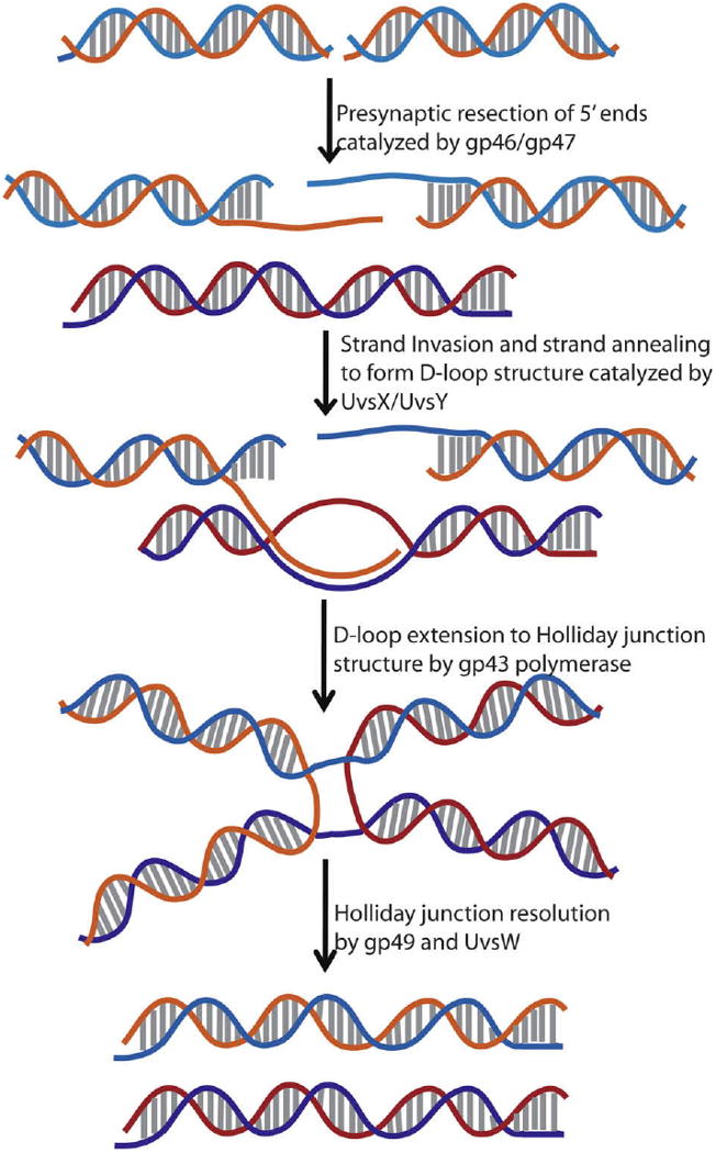
Helicases are an important class of enzymes involved in DNA and RNA metabolism that couple the energy of ATP hydrolysis to unwind duplex DNA and RNA structures. Understanding the mechanism of helicase action is vital due to their involvement in various biological processes such as DNA replication, repair and recombination. Furthermore, the duplex DNA unwinding property of this class of enzymes is closely related to their single-stranded DNA translocation. Hence the study of its translocation properties is essential to understanding helicase activity. Here we review the methods that are employed to analyze the DNA translocation and unwinding activities of the bacteriophage T4 UvsW and Dda helicases. These methods have been successfully employed to study the functions of helicases from large superfamilies.


Figures

Fig. 1
Fig. 1
Schematic representation of the homologous recombination-dependent double strand break (DSB) repair pathway in bacteriophage T4. Gp46 and gp47 complex is involved in the recognition of DSBs and resects 5′-ends to generate 3′-overhangs. These 3′-overhangs are coated with UvsX assisted by UvsY followed by the invasion of a homologous strand to form a D-loop structure. The D-loop is extended by polymerase to generate Holliday junction structures that are resolved by UvsW and gp49 endonuclease, thus producing repaired double strand breaks.
Fig. 2
Fig. 2
Schematic representation of the substrates used in the study of helicases. (A) Blunt ended duplex DNA. (B and C) Partial duplex with 5′ and 3′ ssDNA overhangs, respectively. (D) Bubble DNA structure. (E) Replication fork structure with a primer on the leading strand. (F) Duplex DNA Y substrate. (G) Synthetic Holliday junction structure.
Fig. 3
Fig. 3
Phage T4 UvsW- and Dda-catalyzed unwinding of replication fork, duplex DNA Y substrate and Holliday junction substrates. The reaction products were resolved on a 7.5% PAGE gel shows the products of unwinding. (Reproduced with permission from Ref. [19]).
Fig. 4
Fig. 4
Schematic representation of mobile R-loop and D-loop substrates used in the study of helicases. R-loop structure was synthesized by T7 RNA polymerase, whereas the D-loop structure was produced by RecA mediated strand-invasion reaction.
Fig. 5
Fig. 5
DNA annealing activity mediated by UvsW helicase. A typical time course (0.25–4 min) of ssDNA annealing in the presence of ATP catalyzed by UvsW.
Fig. 6
Fig. 6
2-AP containing replication fork structure with regularly spaced seven 2-AP on the duplex region of the fork. This structure was employed in the fluorescence-based study the unwinding activity of helicase using the fluorescence signal change of 2-AP unwinding the duplex region.
Fig. 7
Fig. 7
Duplex DNA unwinding catalyzed by Dda helicase under pre-steady-state conditions. (A) Products of the helicase reaction showing the separation of the reactants and products on a 20% native PAGE. (B) The data from A with the ssDNA product under pre-steady-state conditions plotted against time. The data were fit to a single exponential followed by a steady-state rate (Eq. (1)). A burst amplitude of 3.5 ± 0.2 nM was obtained when 4 nM Dda helicase was used, implying that the enzyme functions as a monomer during unwinding of the duplex DNA. (Figure reproduced from Ref. [40].)
Fig. 8
Fig. 8
Biotin–streptavidin block assay in the determination of UvsW translocation directionality. (A1 and B1) Schematic representation of the translocation of UvsW helicase on the biotin–streptavidin bearing DNA. Translocation of the enzyme away from the biotin–streptavidin block leads to rapid dissociation from the end of the DNA, whereas dissociation is prevented when translocation occurs in the direction of the block. ATPase activity of UvsW as a function of the 5′-biotin-labeled 30mer (A2) and 3′-biotin-labeled 30mer (B2) in the presence (closed circles) and absence (open circles) of streptavidin (1 μM).
Fig. 9
Fig. 9
(A) Schematic representation of the streptavidin displacement assay of T4 Dda helicase. (B) The products of the assay resolved on a native PAGE. (Reproduced with permission from Ref.[13]).
Scheme 1
Scheme 1
Coupled enzymatic NADH oxidation assay.
Scheme 2
Scheme 2
Kinetic scheme for ATP-dependent ssDNA translocation of UvsW.

Similar articles

Cited by

References

    1. Singleton MR, Dillingham MS, Wigley DB. Annu Rev Biochem. 2007;76:23–50. - PubMed
    1. Ellis NA. Curr Opin Genet Dev. 1997;7:354–363. - PubMed
    1. Nelson SW, Zhuang Z, Spiering MM, Benkovic SJ. T4 Phage Replisome. In: Cameron C, editor. Viral Genome Replication. Springer; New Jersey: 2009. pp. 349–376.
    1. Liu CC, Alberts BM. J Biol Chem. 1981;256:2813–2820. - PubMed
    1. Young MC, Schultz DE, Ring D, von Hippel PH. J Mol Biol. 1994;235:1447–1458. - PubMed

Publication types

LinkOut - more resources