

Recurrent polymorphonuclear pleocytosis with increased red blood cells caused by varicella zoster virus infection of the central nervous system: Case report and review of the literature
- PMID: 20170926
- PMCID: PMC2846975
- DOI: 10.1016/j.jns.2010.01.019
Recurrent polymorphonuclear pleocytosis with increased red blood cells caused by varicella zoster virus infection of the central nervous system: Case report and review of the literature
Abstract
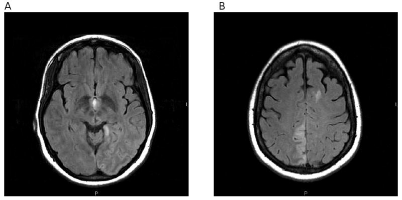
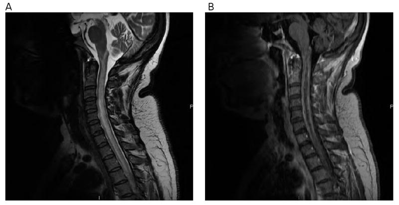
We describe an immunocompetent 45-year-old woman who had four episodes of neurological disease (meningoencephalitis, multifocal vasculopathy, myelitis and inflammatory brain stem disease) produced by varicella zoster virus (VZV) over an 11-month period, all in the absence of rash. The cerebrospinal fluid (CSF) contained anti-VZV IgG antibody, but not VZV DNA throughout her illness, reaffirming the superiority of detection of anti-VZV IgG in CSF compared to VZV DNA in diagnosing VZV infection of the nervous system. Moreover, 3 of 7 CSF samples examined during the 11 months showed a VZV-induced pleocytosis consisting predominantly of polymorphonuclear cells (PMNs), and 4 of 7 samples also contained increased numbers of red blood cells (RBCs). Because increased PMNs and RBCs in CSF can also occur in patients with central and peripheral nervous system disease produced by cytomegalovirus (CMV), the differential diagnosis of chronic nervous system infection with increased PMNs and RBCs in CSF should include analyses for both VZV and CMV.
Copyright 2010 Elsevier B.V. All rights reserved.
Figures
References
-
- Mariotti P, Colosimo C, Frisullo G, Caggiula M, Della Marca GD, Valentini P, et al. Relapsing demyelinating disease after chicken pox in a child. Neurology. 2006;66:1953–4. - PubMed
-
- Gilden DH, Bennett JL, Kleinschmidt-DeMasters BK, Song DD, Yee AS, Steiner I. The value of cerebrospinal fluid antiviral antibody in the diagnosis of neurologic disease produced by varicella zoster virus. J Neurol Sci. 1998;159:140–4. - PubMed
Publication types
MeSH terms
Substances
Grants and funding
LinkOut - more resources
Full Text Sources
Medical
