. 2010 Apr 15;340(2):571-82.
doi: 10.1016/j.ydbio.2010.02.015. Epub 2010 Feb 18.

An efficient approach to isolate STAT regulated enhancers uncovers STAT92E fundamental role in Drosophila tracheal development

Affiliations

An efficient approach to isolate STAT regulated enhancers uncovers STAT92E fundamental role in Drosophila tracheal development

Sol Sotillos et al. Dev Biol. .

Abstract

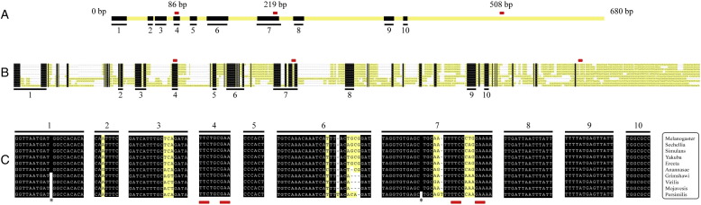
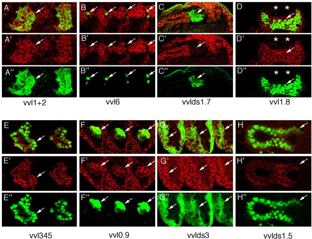
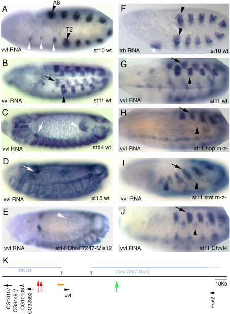
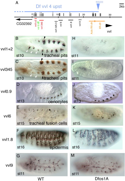
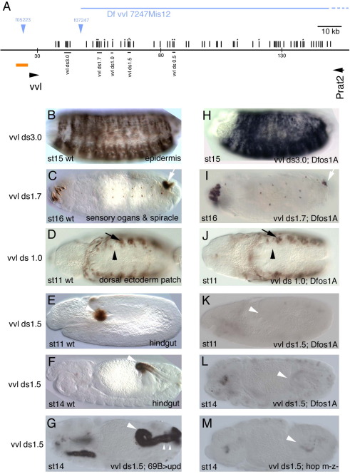
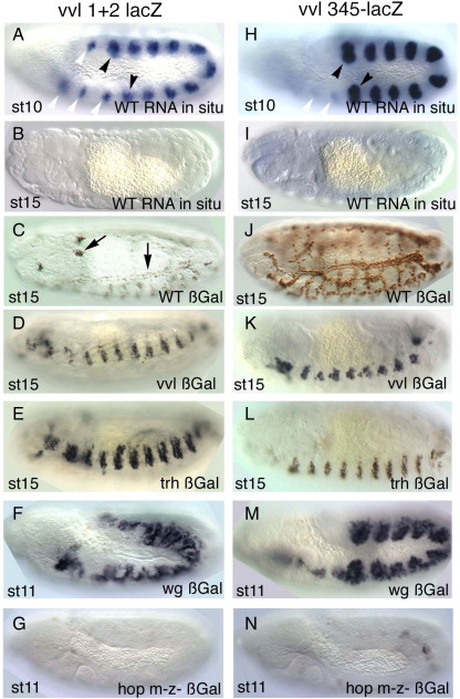
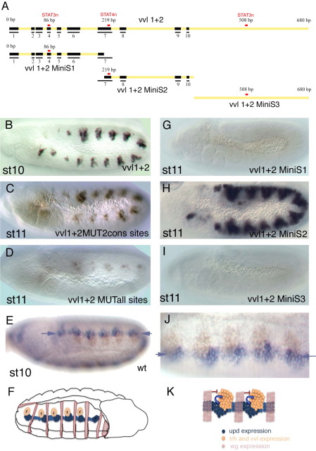
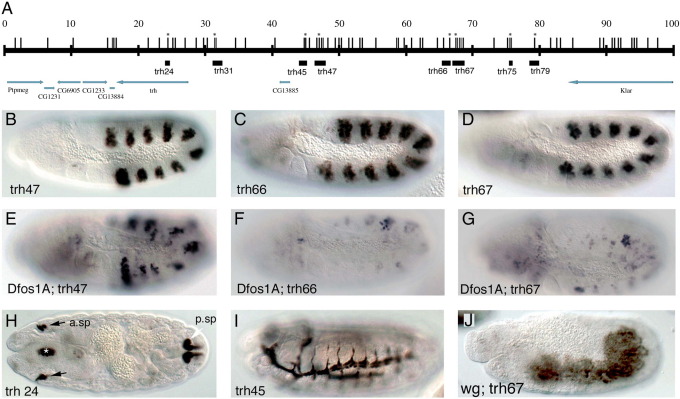
The ventral veinless (vvl) and trachealess (trh) genes are determinants of the Drosophila trachea. Early in development both genes are independently activated in the tracheal primordia by signals that are ill defined. Mutants blocking JAK/STAT signaling at any level do not form a tracheal tree suggesting that STAT92E may be an upstream transcriptional activator of the early trachea determinants. To test this hypothesis we have searched for STAT92E responsive enhancers activating the expression of vvl and trh in the tracheal primordia. We show that STAT92E regulated enhancers can be rapidly and efficiently isolated by focusing the analysis on genomic regions with clusters of putative STAT binding sites where at least some of them are phylogenetically conserved. Detailed analysis of a vvl early tracheal enhancer shows that non-conserved sites collaborate with conserved sites for enhancer activation. We find that STAT92E regulated enhancers can be located as far 60 kb from the promoters. Our results indicate that vvl and trh are independently activated by STAT92E which is the most important transcription factor required for trachea specification.


Figures

Supplementary Fig. 1
Conservation of the vvl1+2 enhancer. (A) Schematic representation of the vvl1+2 enhancer showing as black boxes the regions of sequence conserved in all 10 Drosophila species analyzed. Small red rectangles represent the three D. melanogaster STAT92E sites. Conserved boxes are numbered to relate them to panels B and C. Most homology blocks are grouped in two thirds of the fragment. (B) Alignment of the vvl1+2 fragment in 10 Drosophila species. Black boxes represent regions of absolute sequence conservation, while non-conserved bases are represented in yellow. Note that some of the conserved blocks in D. melanogaster are split in some species and are separated in the figure by white gaps (-). (C) Sequence of the longest conserved blocks shown in A and B. Asterisks mark the location of gaps in some species. Only two STAT92E sites are in conserved sequence blocks. In this area we did not find any other STAT92E sites in other species.
Supplementary Fig. 2
Expression of various enhancers in cells transcribing vvl. Top panels show close ups of double stained embryos of the indicated enhancer constructs labeled with anti-ßGal (green) and vvl RNA in situ (red). Middle panels (′) show vvl RNA. Lower panels (″) show ß-Gal staining. With the exception of vvl 1.7 all enhancers drive expression in cells transcribing vvl. Arrows point to tracheal pits (A and E), isolated trachea cells that could represent fusion cells (B), spiracular chamber cells of the internal posterior spiracle (C), epidermal cells (D and G), oenocytes (F) and posterior hindgut (H). Note in H that both the vvlds1.5 reporter construct and the vvl RNA are expressed in a gradient with lower levels in the posterior hindgut. Panel D is a dorsal view of A8 showing that the posterior spiracles (*) do not express vvl. All other panels show lateral views with anterior left and dorsal up.
Fig. 1
Fig. 1
vvl expression in wild type and mutant backgrounds. All embryos show vvl RNA in situ expression except (F) that shows trh RNA expression. (A–D) Wild type vvl RNA expression. (A) At early st10 vvl is transcribed in the primordia of the 10 tracheal pits (black arrowheads in T2 and A8 mark the first and last primordia) and in homologous cells in segments that will not form trachea (white arrowheads). (B) At st11 10 dorsal epithelial patches (black arrow) appear in each trunk segment displaced with respect to the tracheal primordia (black arrowhead). (C) At st14 epithelial expression has spread to almost all the trunk although certain cells, like some in the posterior spiracles, never express vvl. The anterior hindgut (white arrowhead) also expresses vvl from st11. (D) At st15 high levels of vvl are expressed in the trachea and in a group of cells migrating dorsally (white arrows in C and D) that will join the ring gland. (E) vvl RNA in situ in a Dfvvl 7247-Mis12 embryo lacking most of the vvl downstream region showing absence of hindgut expression (white arrowhead). (F) trh RNA in situ in a wild type st10 embryo showing expression in the salivary gland primordium (asterisk) and in the 10 tracheal pits (black arrowheads mark the first and last pit primordia). (G) Ventro lateral view of a wild type st11 embryo showing vvl expression in the tracheal pit (black arrowhead) and the dorsal patch (arrow). (H) In null hop mutant embryos at st11 vvl is not expressed in the tracheal pits (arrowhead), while the dorsal patches (arrow) and the midline expression develop normally. (I) Similarly, in null stat92E mutant embryos vvl is not expressed at st11 in the tracheal pits (arrowhead), while the dorsal patches (arrow) develop normally. (J) Dfvvl4 embryos with deleted upstream region have no tracheal expression at st10 until at st11 they start developing very weak vvl expression in the tracheal pits (arrowhead). (K) Cartoon depicting the vvl genomic region. The intronless vvl transcript is located at 27 kb from CG32392 and at 117 kb from Prat2. The orange box represents the 7 kb area analyzed in previous works that uncovered the autoregulatory RX-drf enhancer. Red arrows mark the position of the early trachea enhancers, green arrow the position of the hindgut enhancer. Blue triangles represent the FRT piggyBac transposable elements used to delete the upstream and downstream cis-regulatory region (blue lines).
Fig. 2
Fig. 2
Upstream vvl embryonic enhancers in wild type and upd mutant background. (A) Cartoon showing the location of the upstream vvl enhancers analyzed. Vertical lines represent putative STAT92E binding sites. An asterisk marks sites conserved in all 10 studied Drosophilids. Orange line represents the upstream region analyzed previously (Certel et al., 1996). Red lines represent the early tracheal enhancers. Green line represents the oenocyte enhancer. Horizontal blue line indicates the deleted upstream region in Dfvvl4. (B–G) Expression of the indicated enhancers in wild type embryos. (H–M) Expression of the same enhancers in Dfos1A embryos lacking all three upd ligands. Note the strong effect that lack of upd has on vvl1+2 and vvl345 expression (H–I). Black arrowheads point to the tracheal pits, white arrowheads point to the homologous patches in anterior and posterior segments.
Fig. 3
Fig. 3
Downstream vvl embryonic enhancers in wild type and upd mutant backgrounds. (A) Cartoon representing the location of the downstream vvl enhancers analyzed. Symbols are as in Fig. 2. (B–F) Expression of the indicated enhancers in wild type embryos. (H–L) Expression of the same enhancers in a Dfos1A embryos lacking all three upd ligands. (K–L) Note the strong effect that lack of upd has on vvlds1.5 hindgut expression. (G) Ectopic upd results in ectopic vvlds1.5 expression in the most posterior area of the hindgut (small arrowheads) and in the salivary glands. (M) In null hop mutant embryos vvlds1.5 is not expressed in the hindgut (arrowhead).
Fig. 4
Fig. 4
Expression of early vvl tracheal enhancers in various mutant backgrounds. (A–G) vvl1+2 expression and (H–N) vvl345 expression. (A) lacZ RNA in situ of a wild type vvl1+2 st10 embryo showing the expression in the tracheal primordia (black arrowheads point the primordia in T2 and in A8) and in the anterior and posterior spots that will not form trachea (white arrowheads). (B) lacZ RNA in situ of a vvl1+2 st15 embryo showing the enhancer is not expressed at later stages. (C) ß-Gal expression of a vvl1+2 st15 wild type embryo. Note that long-lived ß-Gal perduring expression can be still detected in the trachea and in a group of cells moving dorsally (Arrows). (D) In vvl mutants vvl1+2 is activated but the expressing cells do not form normal trachea. (E) In trh mutant embryos ß-Gal expressing cells do not invaginate staying on the embryo's epidermal surface. (F) In wg mutant embryos vvl1+2 expression is not restricted to the tracheal primordia. Note that levels of expression are still lower outside the normal pit position. (G) vvl1+2 expression is almost missing in hop mutants lacking maternal and zygotic JAK. (H) lacZ RNA in situ of a vvl345 st10 embryo showing the expression in the tracheal primordia (black arrowheads point the primordia in T2 and in A8). The expression in the homologous spots in anterior segments is very weak (white arrowheads). (I) lacZ RNA in situ of a vvl345 st15 embryo showing that the enhancer is not expressed any longer. (J) ß-Gal expression of a vvl345 st15 embryo. The perduring protein can be detected by the antibody. (K) In vvl mutants vvl345 is activated but the invaginated cells do not fuse into a tracheal tree. (L) In trh mutants vvl345 is expressed but the cells remain on the embryonic epidermis. (M) In wg mutants vvl345 expression is not restricted to the tracheal pits. (N) vvl345 expression is almost missing in hop mutants lacking maternal and zygotic JAK. (A–B, H–I) show lacZ RNA in situ. (C–G, J–N) show anti-ßGal.
Fig. 5
Fig. 5
Expression of the vvl1+2 early tracheal enhancers is controlled by STAT92E signaling. (A) Scheme of vvl1+2 and subfragments tested. Black boxes represent blocks of conserved sequence in all 10 Drosophila species analyzed (see supplementary Fig.1 for sequence). Red rectangles represent the location of the three putative STAT92E DNA-binding sites. Only two of the sites are located in conserved blocks while the third one is only present in D. melanogaster. (B) Wild type vvl 1+2 expression at st10. (C) ß-Gal expression driven from a vvl 1+2 construct where the two conserved putative STAT92E binding sites have been mutated. Expression is lower than in the wild type, especially anterior to the second abdominal segment, but substantial levels of expression remain. (D) ß-Gal expression driven from a vvl 1+2 construct with all three putative STAT92E binding sites mutated. Expression is highly reduced but traces still remain. (G) The vvl1+2 MiniS1 fragment does not drive tracheal expression. (H) vvl MiniS2 drives tracheal expression in embryos at st11. The expression seems to be less restricted with some ectopic signal between the pits. (I) The vvl1+2 MiniS3 fragment does not drive tracheal expression. (E, J) A wild type st10 embryo double stained to show the upd RNA expression (purple) and the vvl RNA expression (brown). (J) is a magnification of E that has been rotated 180 degrees to have dorsal up and anterior left. Note the continuous upd stripe (purple arrows) running along the antero-posterior axis of the embryo just ventral to the vvl tracheal expression. (F) Scheme of the embryo shown in E that has been rotated 180° like panel J to have dorsal up and anterior left in the region of interest. Besides the tracheal pits and upd expression the scheme shows the location of the wg expressing cell stripes. (K) Scheme of proposed negative and positive regulation of early enhancers in the tracheal placodes. (B–D, G–I) Embryos are stained with anti-ßGal antibody. (E, J) show upd RNA in purple and vvl RNA in brown.
Fig. 6
Fig. 6
trh embryonic enhancers in wild type and mutant backgrounds. (A) Cartoon representing the location of the trh enhancers analyzed. Vertical lines represent putative STAT92E binding sites. An asterisk marks sites conserved in all Drosophilids. Black boxes represent the analyzed regions. Arrows indicate transcribed regions. Note that CG13885 nests inside the trh cis-regulatory region. (B–D) Expression of three early trh tracheal enhancers in a wild type background. (E–G) Expression in Dfos1A embryos of the same enhancers shown in (B–D). Most expression disappears from trh66 and trh67 while substantial signal is still present in trh47. (H) Posterior (white arrow) and anterior spiracle (black arrows) expression driven by trh24, there is also some expression in the pharyngeal ectoderm (asterisk). (I) Expression of the trh45 late trachea enhancer. (J) Expression of trh67 in a wingless mutant background. Lateral views with head left dorsal up in all embryos except H that shows a frontal section.
Fig. 7
Fig. 7
Expression of early trh tracheal enhancers in various mutants. (A–C) ß-Gal expression of the early enhancers at st13, all lines delineate the internalized tracheal network. (D–F) Expression of the early trh trachea enhancers is not affected in homozygous vvl mutant embryos despite the aberrant migration of the tracheal cells. (G–H) Expression of the trh47 and trh66 is maintained in trh mutant embryos with the uninvaginated ectodermal cells remaining on the embryo surface. (I) Expression of the trh67 enhancer requires trh function suggesting a strong degree of autoregulation. (J–L) Expression of trh47 and trh66 in double vvl trh mutant embryos is maintained, while trh67 is absent. Embryos in panels A–I are homozygous for the reporter constructs while in panels J–L are heterozygous.

Similar articles

Cited by

References

    1. Anderson M.G., Certel S.J., Certel K., Lee T., Montell D.J., Johnson W.A. Function of the Drosophila POU domain transcription factor drifter as an upstream regulator of breathless receptor tyrosine kinase expression in developing trachea. Development. 1996;122:4169–4178. - PubMed
    1. Anderson M.G., Perkins G.L., Chittick P., Shrigley R.J., Johnson W.A. drifter, a Drosophila POU-domain transcription factor, is required for correct differentiation and migration of tracheal cells and midline glia. Genes Dev. 1995;9:123–137. - PubMed
    1. Arbouzova N.I., Zeidler M.P. JAK/STAT signalling in Drosophila: insights into conserved regulatory and cellular functions. Development. 2006;133:2605–2616. - PubMed
    1. Baeg G.H., Zhou R., Perrimon N. Genome-wide RNAi analysis of JAK/STAT signaling components in Drosophila. Genes Dev. 2005;19:1861–1870. - PMC - PubMed
    1. Bergad P.L., Shih H.M., Towle H.C., Schwarzenberg S.J., Berry S.A. Growth hormone induction of hepatic serine protease inhibitor 2.1 transcription is mediated by a Stat5-related factor binding synergistically to two gamma-activated sites. J. Biol. Chem. 1995;270:24903–24910. - PubMed

Publication types

MeSH terms

Substances