. 2010 Jul;34(7):925-31.
doi: 10.1016/j.leukres.2010.01.020. Epub 2010 Feb 19.

Arsenic trioxide and ascorbic acid demonstrate promising activity against primary human CLL cells in vitro

Affiliations

Arsenic trioxide and ascorbic acid demonstrate promising activity against primary human CLL cells in vitro

Sabyasachi Biswas et al. Leuk Res. 2010 Jul.

Abstract

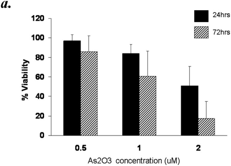
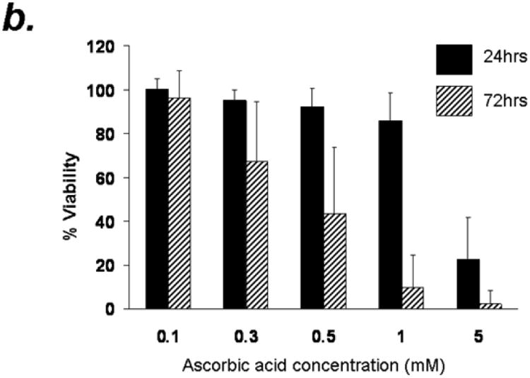
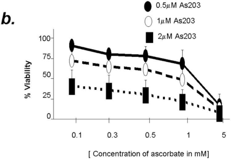
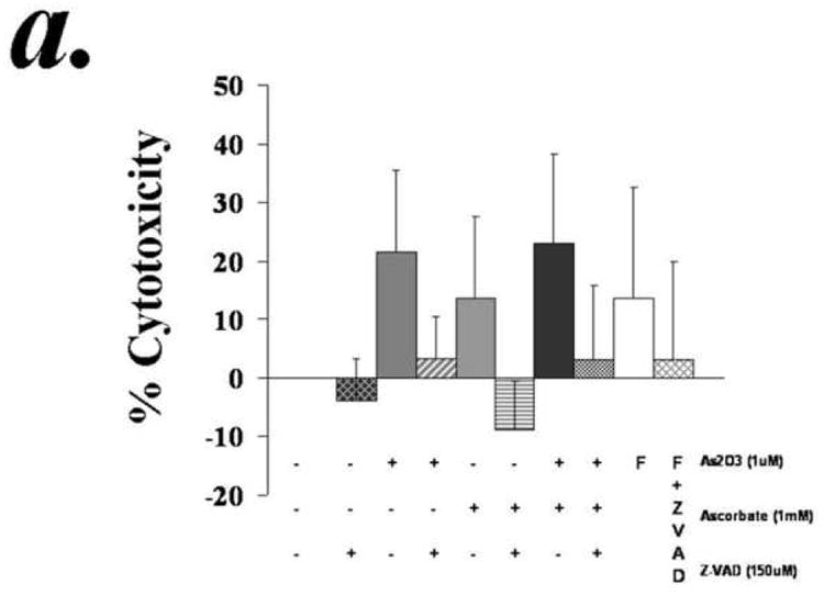
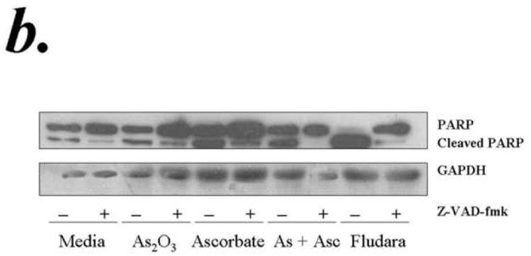
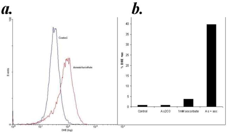
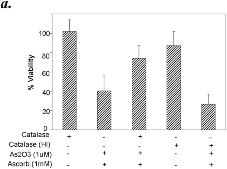
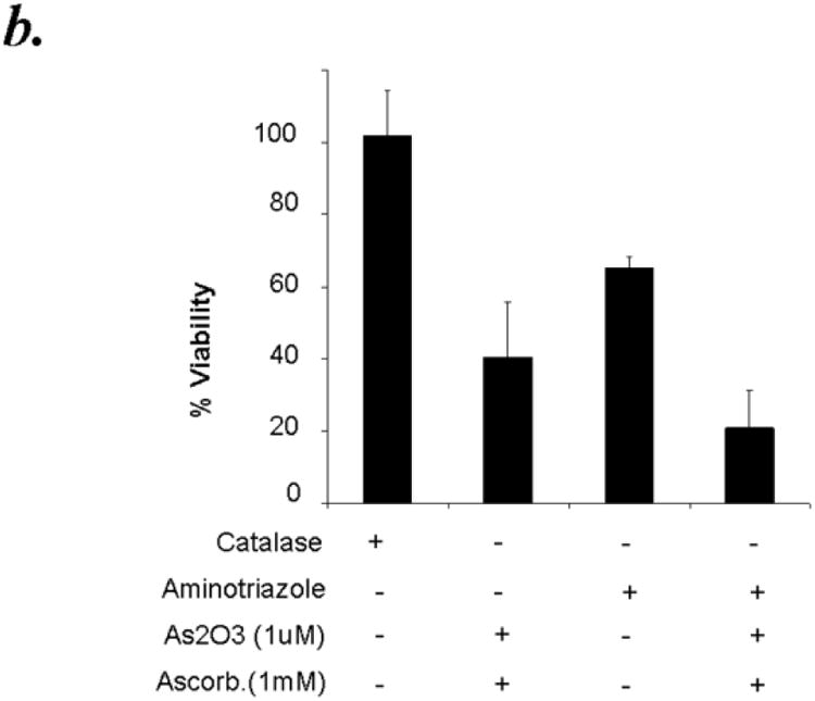
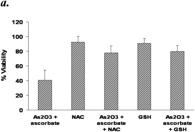
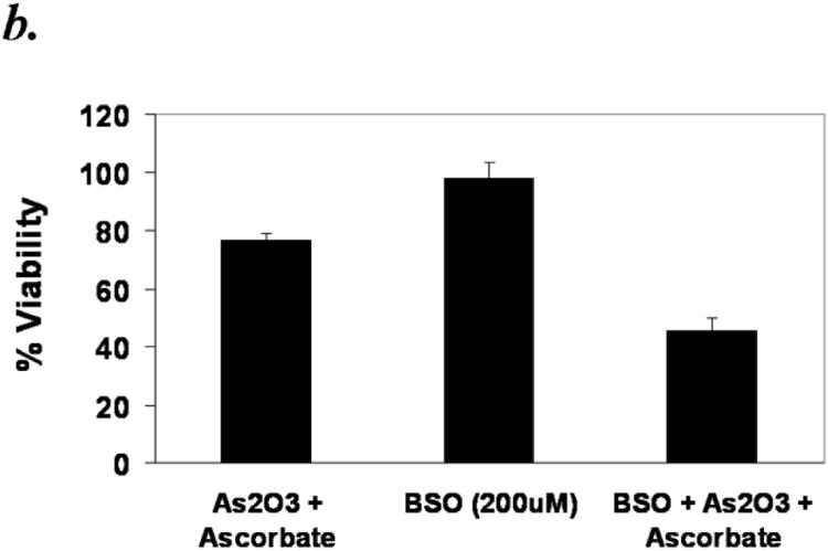
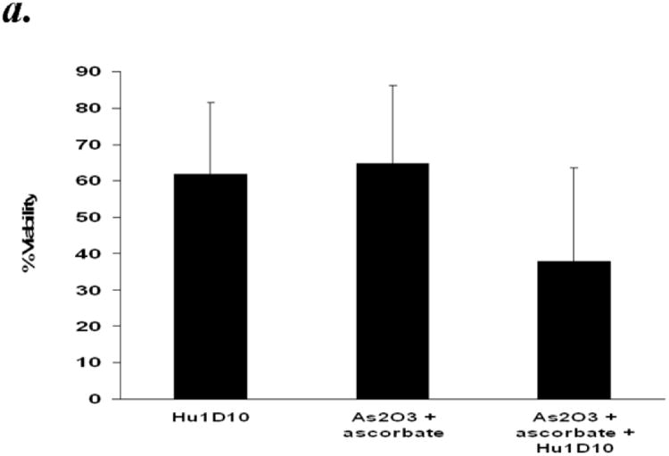
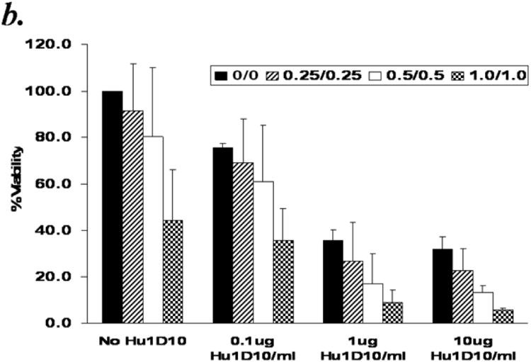
The compromised antioxidant defense system in chronic lymphocytic leukemia (CLL) suggested a potential use for reactive oxygen species (ROS) generating arsenic trioxide (ATO) and ascorbic acid. While both ATO and ascorbic acid mediate cytotoxicity in CLL B cells as single agents, the efficacy of ATO is enhanced by ascorbic acid. This effect is dependent on increased ROS accumulation, as pretreatment of B-CLL cells with a glutathione reducing buthionine sulfoximine or catalase inhibiting aminotriazole, enhanced ATO/ascorbic acid-mediated cytotoxicity. Pretreatment with reducing agents such as catalase, or thiol antioxidant, N-acetyl cysteine or GSH also abrogated ATO/ascorbic acid-mediated cytotoxicity. Furthermore, Hu1D10-mediated cell death was enhanced with ATO and ascorbic acid, thus justifying potential combination of ATO/arsenic trioxide therapy with antibodies such as Hu1D10 that also cause accumulation of ROS.


Conflict of interest statement

Conflict of Interest: The authors declare no conflict of interest.

Figures

Figure 1
Figure 1. Arsenic trioxide or ascorbic acid mediated cytotoxicity in B cells from B-CLL patients- Dose and time kinetic analysis
Purified B-lymphocytes (1 × 10⁶cells/ml) from B-CLL patients were incubated in the presence of indicated concentrations of arsenic trioxide (Panel A) or ascorbic acid (Panel B) for indicated time periods. The cells were stained with Annexin-V-FITC and propidium iodide as described by us previously. The cells were analyzed by flow cytometry and data collected under list mode. The data shown represent % Annexin-V-/PI- viable cells ± SD that are normalized to media control. (n=10)
Figure 2
Figure 2. Combination of Arsenic trioxide and ascorbic acid enhance cytotoxicity in B cells from B-CLL patients
Panel a: Purified B-lymphocytes from CLL patients (1×10⁶/ml media) were incubated with either arsenic trioxide [ATO] (1μM), ascorbic acid (1mM), or with both agents together for the indicated time periods. The cells were stained with Annexin-V-FITC and propidium iodide and analyzed by flow cytometry and data collected under list mode. The data shown represent % Annexin-V-/PI- viable cells ± SD that are normalized to media control. (n=12) Panel b: Purified B-lymphocytes from CLL patients (1×10⁶/ml media) were incubated with 0.5, 1 and 2μM arsenic trioxide [ATO] in conjunction with indicated concentrations of ascorbic acid for 24 hours. The cells were stained with Annexin-V-FITC and propidium iodide and analyzed by flow cytometry and data collected under list mode. The data shown represent % Annexin-V-/PI- viable cells ± SD that are normalized to media control. (n=12; p<0.001).
Figure 3
Figure 3. Arsenic trioxide and ascorbic acid induced cytotoxicity in CLL cells is dependent on activation of caspase
Purified B-lymphocytes from CLL patients (1×10⁶/ml media) were incubated with 1μM arsenic and 1mM ascorbic acid either alone or in combination in the presence or absence of z-VAD-fmk (150uM) for 24 hours. The cells were stained with Annexin-V-FITC and propidium iodide and analyzed by flow cytometry for apoptotic cells (panel a). The data shown represent % Annexin-V+/PI+ cells ± SD.(n=3). Panel B shows Westernblot analysis of the lysates from one of the above experiments was assessed for incleaved and cleaved PARP. Fludarabin is used as a control in these studies.
Figure 4
Figure 4. Accumulation of reactive oxygen species by arsenic trioxide and ascorbic acid
Purified B-lymphocytes from CLL patients (1×10⁶/ml media) (1×10⁶/ml) were treated with 1μM arsenic and 1mM ascorbic acid either alone or in combination. Subsequently DHE was added to 10μM final concentration and the cells were incubated for an additional 60 minutes. The cells were analyzed by flow cytometry and data collected under list mode. A representative histogram showing the vehicle control and arsenic trioxide [ATO]/ascorbic acid treated group is shown in the top panel. The bottom panel shows %DHE positive cells in media control, ATO (0.1uM), ascorbic acid (1mM) and ATO/ascorbic acid treated groups.
Figure 5
Figure 5
Panel a: Active but not heat inactivated catalase, protects against As2O3 and ascorbic acid mediated cytotoxicity in primary CLL B cells. Purified B-lymphocytes from CLL patients (1×10⁶/ml media) were pretreated with 500 units of active catalase or heat inactivated catalase (HI) for 30 minutes prior to addition of arsenic trioxide [ATO] (1μM) and ascorbic acid (1mM). The cells were stained with Annexin-V-FITC and propidium iodide and the cells were analyzed by flow cytometry after 24 hours. The data shown represent % Annexin-V-/PI- viable cells ± SD that are normalized to media control. (n=6; p<0.05). Panel b: Catalase inhibitor aminotriazole enhances the cytotoxicity of As2O3 and ascorbic acid towards primary CLL B cells. Purified B-lymphocytes from CLL patients (1×10⁶/ml media) were pretreated with 100mM aminotriazole for 30 minutes prior to addition of arsenic trioxide [ATO] (1μM) and ascorbic acid (1mM). The cells were stained with Annexin-V-FITC and propidium iodide and the cells were analyzed by flow cytometry after 24 hours. The data shown represent % Annexin-V-/PI-viable cells ± SD that are normalized to media control. (n=6; p<0.05).
Figure 5
Figure 5
Panel a: Active but not heat inactivated catalase, protects against As2O3 and ascorbic acid mediated cytotoxicity in primary CLL B cells. Purified B-lymphocytes from CLL patients (1×10⁶/ml media) were pretreated with 500 units of active catalase or heat inactivated catalase (HI) for 30 minutes prior to addition of arsenic trioxide [ATO] (1μM) and ascorbic acid (1mM). The cells were stained with Annexin-V-FITC and propidium iodide and the cells were analyzed by flow cytometry after 24 hours. The data shown represent % Annexin-V-/PI- viable cells ± SD that are normalized to media control. (n=6; p<0.05). Panel b: Catalase inhibitor aminotriazole enhances the cytotoxicity of As2O3 and ascorbic acid towards primary CLL B cells. Purified B-lymphocytes from CLL patients (1×10⁶/ml media) were pretreated with 100mM aminotriazole for 30 minutes prior to addition of arsenic trioxide [ATO] (1μM) and ascorbic acid (1mM). The cells were stained with Annexin-V-FITC and propidium iodide and the cells were analyzed by flow cytometry after 24 hours. The data shown represent % Annexin-V-/PI-viable cells ± SD that are normalized to media control. (n=6; p<0.05).
Figure 6
Figure 6
Panel a: N-acetyl cysteine (NAC) or Glutathione (GSH) mediated abrogation arsenic trioxide/ascorbic acid induced cytotoxicity in primary CLL B cells. Purified B-lymphocytes from CLL patients (1×10⁶/ml media) were pretreated with Glutathione [GSH (1mM)] or N-acetyl cysteine (NAC) for 30 minutes prior to addition of ATO (1μM) and ascorbic acid (1mM). The cells were stained with Annexin-V-FITC and propidium iodide and analyzed by flow cytometry after 24 hours. The data shown represent % Annexin-V-/PI- viable cells + SD that are normalized to media control. (n=6; p<0.05). Panel b: Depletion of GSH by buthionine sulfoximine (BSO) enhances the As2O3 and ascorbic acid mediated cytotoxicity. Purified CLL B cells (1×10⁶/ml) were grown in the presence of 200μM BSO for 24 hours to deplete GSH levels. The cells were analyzed by flow-cytometry 24 hours after addition of ATO/ascorbic acid as described above.
Figure 6
Figure 6
Panel a: N-acetyl cysteine (NAC) or Glutathione (GSH) mediated abrogation arsenic trioxide/ascorbic acid induced cytotoxicity in primary CLL B cells. Purified B-lymphocytes from CLL patients (1×10⁶/ml media) were pretreated with Glutathione [GSH (1mM)] or N-acetyl cysteine (NAC) for 30 minutes prior to addition of ATO (1μM) and ascorbic acid (1mM). The cells were stained with Annexin-V-FITC and propidium iodide and analyzed by flow cytometry after 24 hours. The data shown represent % Annexin-V-/PI- viable cells + SD that are normalized to media control. (n=6; p<0.05). Panel b: Depletion of GSH by buthionine sulfoximine (BSO) enhances the As2O3 and ascorbic acid mediated cytotoxicity. Purified CLL B cells (1×10⁶/ml) were grown in the presence of 200μM BSO for 24 hours to deplete GSH levels. The cells were analyzed by flow-cytometry 24 hours after addition of ATO/ascorbic acid as described above.
Figure 7
Figure 7
Panel a: Arsenic trioxide and ascorbic acid enhance Hu1D10 mediated cytotoxicity of primary CLL B cells. Purified B-lymphocytes from CLL patients (1×10⁶/ml media) were treated with Hu1D10 (10ug/ml), arsenic trioxide [ATO](1μM)/ascorbic acid (1mM) or Hu1D10 and ATO/ascorbic acid. The cells were stained with Annexin-V-FITC and propidium iodide and analyzed by flow cytometry as described above. The data shown represent % Annexin-V-/PI- viable cells ± SD that are normalized to media control. (n=11). Panel b: Arsenic trioxide and ascorbic acid enhance the cytotoxicity of Hu1D10 in a dose dependent manner. Purified B-lymphocytes from CLL patients (1×10⁶/ml media) were treated with indicated concentrations of Hu1D10, arsenic trioxide [ATO] and ascorbic acid. The cells were stained with Annexin-V-FITC and propidium iodide and analyzed by flow cytometry as described above. The data shown represent % Annexin-V-/PI- viable cells ± SD that are normalized to media control. Varying arsenic trioxide/ascorbic acid and Hu1D10 concentrations show that even if Hu1D10 concentration is lowered 10 fold, the cytotoxicity in conjunction with ATO/ascorbic acid is significantly enhanced.
Figure 7
Figure 7
Panel a: Arsenic trioxide and ascorbic acid enhance Hu1D10 mediated cytotoxicity of primary CLL B cells. Purified B-lymphocytes from CLL patients (1×10⁶/ml media) were treated with Hu1D10 (10ug/ml), arsenic trioxide [ATO](1μM)/ascorbic acid (1mM) or Hu1D10 and ATO/ascorbic acid. The cells were stained with Annexin-V-FITC and propidium iodide and analyzed by flow cytometry as described above. The data shown represent % Annexin-V-/PI- viable cells ± SD that are normalized to media control. (n=11). Panel b: Arsenic trioxide and ascorbic acid enhance the cytotoxicity of Hu1D10 in a dose dependent manner. Purified B-lymphocytes from CLL patients (1×10⁶/ml media) were treated with indicated concentrations of Hu1D10, arsenic trioxide [ATO] and ascorbic acid. The cells were stained with Annexin-V-FITC and propidium iodide and analyzed by flow cytometry as described above. The data shown represent % Annexin-V-/PI- viable cells ± SD that are normalized to media control. Varying arsenic trioxide/ascorbic acid and Hu1D10 concentrations show that even if Hu1D10 concentration is lowered 10 fold, the cytotoxicity in conjunction with ATO/ascorbic acid is significantly enhanced.

Similar articles

Cited by

References

    1. Farber CM, Liebes LF, Kanganis DN, Silber R. Human B lymphocytes show greater susceptibility to H2O2 toxicity than T lymphocytes. J Immunol. 1984;132:2543–2546. - PubMed
    1. Oltra AM, Carbonell F, Tormos C, Iradi A, Saez GT. Antioxidant enzyme activities and the production of MDA and 8-oxo-dG in chronic lymphocytic leukemia. Free Radic Biol Med. 2001;30:1286–1292. - PubMed
    1. Farber CM, Kanganis DN, Liebes LF, Silber R. Antioxidant enzymes in lymphocytes from normal subjects and patients with chronic lymphocytic leukaemia: increased glutathione peroxidase activity in CLL B lymphocytes. Br J Haematol. 1989;72:32–35. - PubMed
    1. Moran EC, Kamiguti AS, Cawley JC, Pettitt AR. Cytoprotective antioxidant activity of serum albumin and autocrine catalase in chronic lymphocytic leukaemia. Br J Haematol. 2002;116:316–328. - PubMed
    1. Ivanov VN, Hei TK. Arsenite sensitizes human melanomas to apoptosis via tumor necrosis factor alpha-mediated pathway. J Biol Chem. 2004;279:22747–22758. - PMC - PubMed

Publication types

MeSH terms