. 2010 Mar 26;394(1):12-7.
doi: 10.1016/j.bbrc.2010.02.112. Epub 2010 Feb 19.

FSH-receptor isoforms and FSH-dependent gene transcription in human monocytes and osteoclasts

Affiliations

FSH-receptor isoforms and FSH-dependent gene transcription in human monocytes and osteoclasts

Lisa J Robinson et al. Biochem Biophys Res Commun. .

Abstract

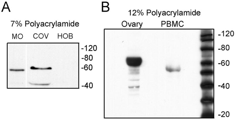
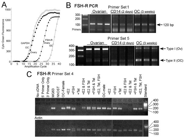
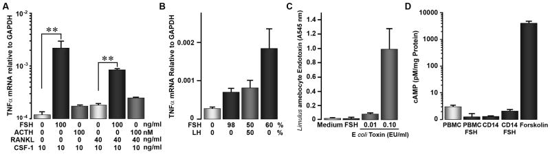
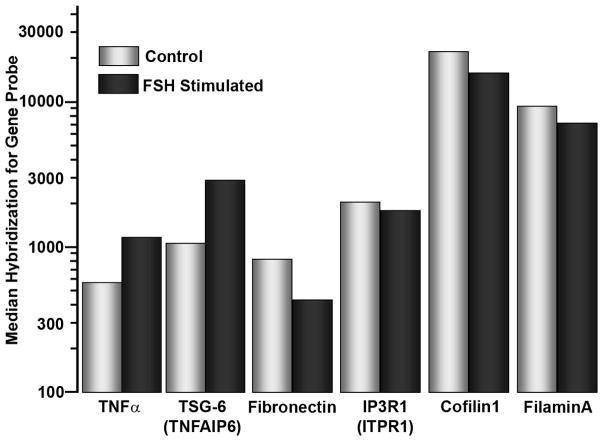
Cells of the monocyte series respond to follicle stimulating hormone (FSH) by poorly characterized mechanisms. We studied FSH-receptors (FSH-R) and FSH response in nontransformed human monocytes and in osteoclasts differentiated from these cells. Western blot and PCR confirmed FSH-R expression on monocytes or osteoclasts, although at low levels relative to ovarian controls. Monocyte and osteoclast FSH-Rs differed from FSH-R from ovarian cells, reflecting variable splicing in exons 8-10. Monocytes produced no cAMP, the major signal in ovarian cells, in response to FSH. However, monocytes and osteoclasts transcribed TNFalpha in response to the FSH. No relation of expression of osteoclast FSH-R to the sex of cell donors or to exposure to sex hormones was apparent. Controls for FSH purity and endotoxin contamination were negative. Unamplified cRNA screening in adherent CD14 cells after 2h in 25ng/ml FSH showed increased transcription of RANKL signalling proteins. Transcription of key proteins that stimulate bone turnover, TNFalpha and TSG-6, increased 2- to 3-fold after FSH treatment. Smaller but significant changes occurred in transcripts of selected signalling, adhesion, and cytoskeletal proteins. We conclude that monocyte and osteoclast FSH response diverges from that of ovarian cells, reflecting, at least in part, varying FSH-R isoforms.


Figures

Figure 1
Figure 1. Western blots of human peripheral blood monocytes for FSH receptor
A. Western blot of 20 μg cell extracts separated on 7% acrylamide analyzed using anti FSH-R (AbCam) recognizing an internal peptide of the receptor. Left: human monocytes (MO) in RANKL and CSF-1 3 days. Middle: ovarian cell extract (COV; positive control). Right: human osteoblasts (negative). Irrelevant lanes between MO and COV are deleted for better comparison of Mr. B. High resolution Western blot. Fifty μg cell extracts separated on 12% polyacrylamide were evaluated using antibody (Santa Cruz) to an N-terminal epitope of the FSH-R. A protein with Mr ~55 kD is again identified in isolated human peripheral blood monocytes (PBMC). The positive control (ovarian extract, left) has much more FSH-R than PBMC, as in (A), with Mr ~ 60 kD.
Fig 2
Fig 2. FSH-R in human monocytes and osteoclasts by PCR assays
All FSH-R amplicons cross exons to avoid amplifying trace genomic DNA (see Methods) A. GAPDH and FSH-R signal indicated as fluorescence of Sybr green (abscissa) as a function of cycle number (ordinate). The data use cDNA samples amplified with FSH-R primer set 1; the amplified FSH-R fragments (right two curves) are shown in the top row of (B), lanes 3 and 9. B. FSH-R transcripts produced by monocytes and osteoclasts mainly exclude exon 9 of ovarian FSH-R. Probe-set 1 (top panel) shows results typical of probes including an intron but in regions without known variability in mRNA. The product in three independent reactions using CD14 cells was slightly less than in osteoclasts, and much less than in ovarian cells (see A). Probe-sets that amplify variants of the receptor including or excluding exon 9 differentiated FSH-R in the different cell types (bottom). The major amplicon in ovarian cells is the type 1 transcript containing exons 8, 9, and 10 (320 bp band). A trace of the type 2 transcript (140 bp) was also present, in keeping with other reports. In contrast, assays using CD14 macrophages or osteoclasts after three weeks in CSF-1 and RANKL showed major isoforms with sizes of ~140 bp but only traces of the ovarian form containing exon 9. Replicates are all from independent PCR reactions. Products were verified by sequencing (see text). C. FSH-R expression in osteoclasts does not vary with cell donor sex or with exposure to FSH or sex steroids. No consistent difference in FSH-receptor was seen in cells in osteoclast differentiation medium 10 days using monocytes from male or female donors, with or without sex steroids and FSH. Additions as indicated were: 10 nM estradiol (E2), 100 ng/ml of FSH, or 10 nM Delatestryl (Tst). Amplification of FSH-R (top panel) used primer pair 4. The 142 bp region amplified in all but three osteoclast samples; negatives occurred randomly and probably reflect technical errors. The split bands in a few samples are believed to be artifacts but were not in this case sequenced. Controls shown are actin amplified from the same mRNA samples (bottom panel) and FSH-R reactions from human osteoblast (NhOst) cDNA (negative), and the osteoblast-like cell line MG63, in which detectible FSH-R was present.
Figure 3
Figure 3. FSH increases FSH mRNA in adherent human monocytes
There was variation in quantitative response between assays and between FSH preparations, but all cell preparations and all FSH sources gave qualitatively similar effects. A. In 7 day cultures of adherent monocytes and osteoclasts, production of TNFα mRNA by real-time PCR was increased by addition of 25 ng/ml FSH. ACTH, 100 nM, is included as a control. Medium was Dubelco’s MEM with 10% fetal calf serum and 10 ng/ml CSF-1; RANKL for osteoclast differentiation was 40 ng/ml. N=2., mean ± range. B. Different sources of FSH increase TNFα production; admixture with LH had no effect. TNFα mRNA transcription in response to three preparations of human FSH are shown. Highly pure and partially pure FSH caused significant increases in TNFα mRNA relative to controls without FSH (left bar) A mixture of pure FSH with an equal amount LH (third bar), did not change response relative to FSH alone. N=2, mean ± range. C. Endotoxin does not contribute to the FSH effect. The Limulus amebocyte assay was used; endotoxin controls at 0.01 and 0.1 EU/ml are shown (right two bars). Osteoclast medium and each FSH sample gave results corresponding to endotoxin levels of 0.001 EU/ml or less. N=2, mean ± range. D. Cyclic AMP is not produced in response to FSH, 25 ng/ml, 15 minutes, added to mononuclear cell cultures. A standard curve was used to calculate cAMP. Control untreated and FSH treated mononuclear cells and osteoclasts are shown, in addition to a forskolin (1 μM, 15 minutes) positive control cells (right bar). Note the logarithmic scale.
Figure 4
Figure 4. Changes in selected genes in two day CD14 adherent monocytes after two hour FSH treatment (25 ng/ml)
The ordinate roughly correlates with transcript abundance. The data show 2–3 fold (note log scale) for increases in TNFα and TSG-6 mRNA after FSH treatment, and ~2-fold decrease in fibronectin. Cytoskeletal organizing proteins, including cofilin1 and filaminA, were modestly, ~40%, but consistently decreased. Multiple probes showed similar magnitude effects, except for TSG-6, for which only one probe-set was included in the array. For each measurement, n=20. All differences shown had p < 0.001 between untreated and FSH treated measurements in each pair.

References

    1. Marians RC, Ng L, Blair HC, et al. Defining thyrotropin-dependent and-independent steps of thyroid hormone synthesis by using thyrotropin receptor-null mice. Proc Natl Acad Sci USA. 2002;99:15776–15781. - PMC - PubMed
    1. Abe E, Marians RC, Yu W, et al. TSH is a negative regulator of skeletal remodeling. Cell. 2003;115:151–162. - PubMed
    1. Sun L, Sharrow AC, Zhang Z, et al. FSH Directly Regulates Bone Mass. Cell. 2006;125:247–260. - PubMed
    1. Iqbal J, Sun L, Blair HC, Zaidi M. FSH Stimulates TNF Production From Immune Cells. Proc Natl Acad Sci USA. 2006;103:14925–14930. - PMC - PubMed
    1. Yee JB, Hutson JC. Testicular macrophages: isolation, characterization and hormonal responsiveness. Biol Reprod. 1983;29:1319–1326. - PubMed

MeSH terms