

Functional analysis of Scr during embryonic and post-embryonic development in the cockroach, Periplaneta americana
- PMID: 20171962
- PMCID: PMC2856087
- DOI: 10.1016/j.ydbio.2010.02.018
Functional analysis of Scr during embryonic and post-embryonic development in the cockroach, Periplaneta americana
Abstract
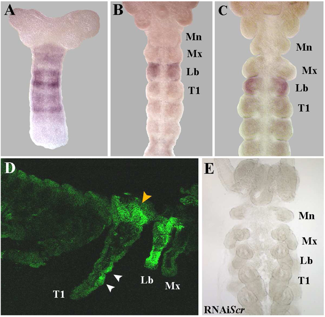
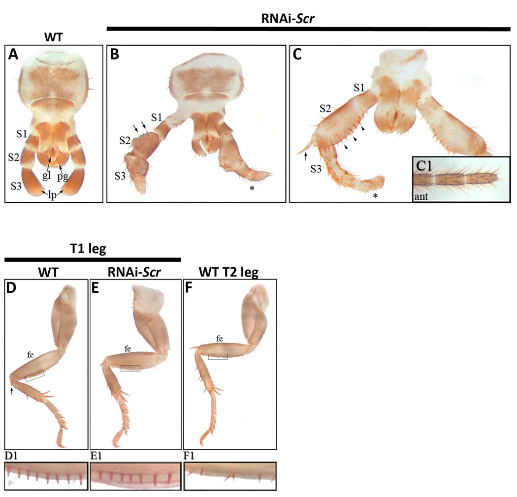
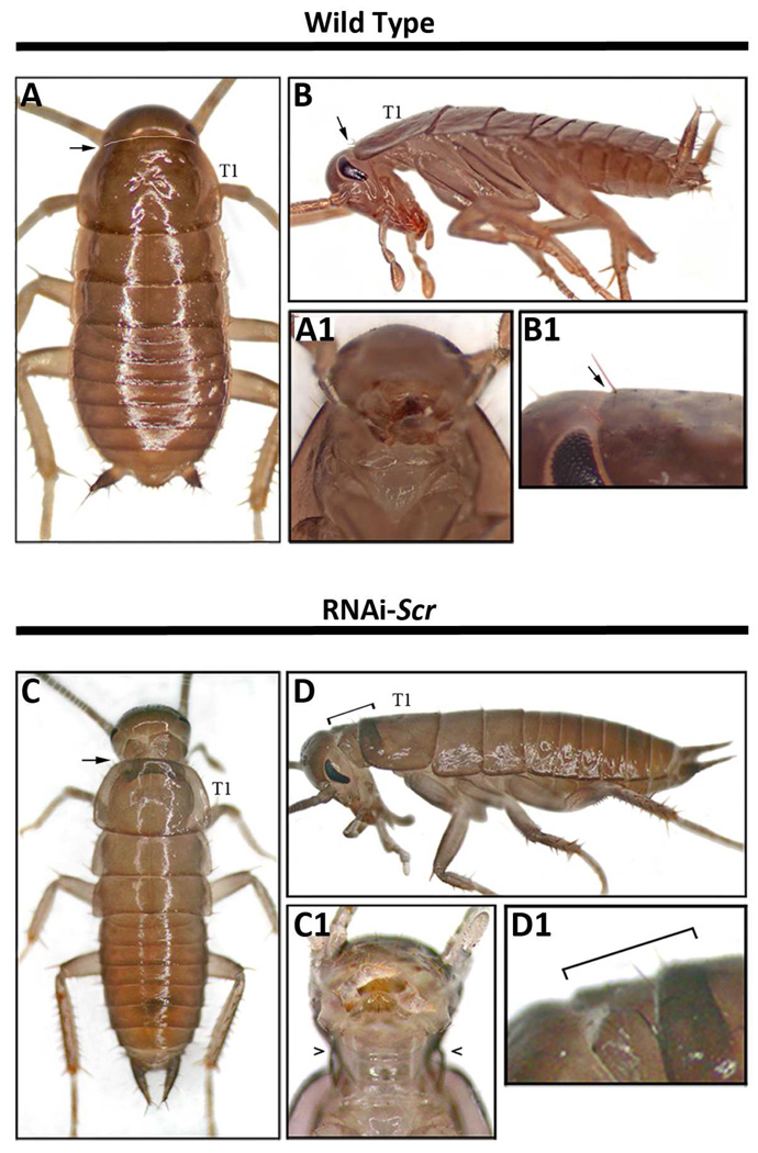
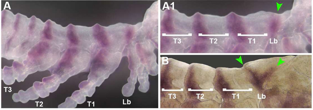
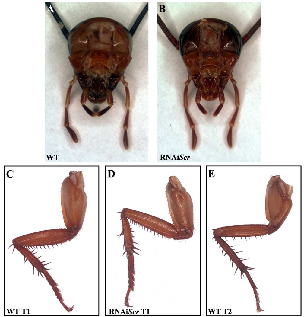
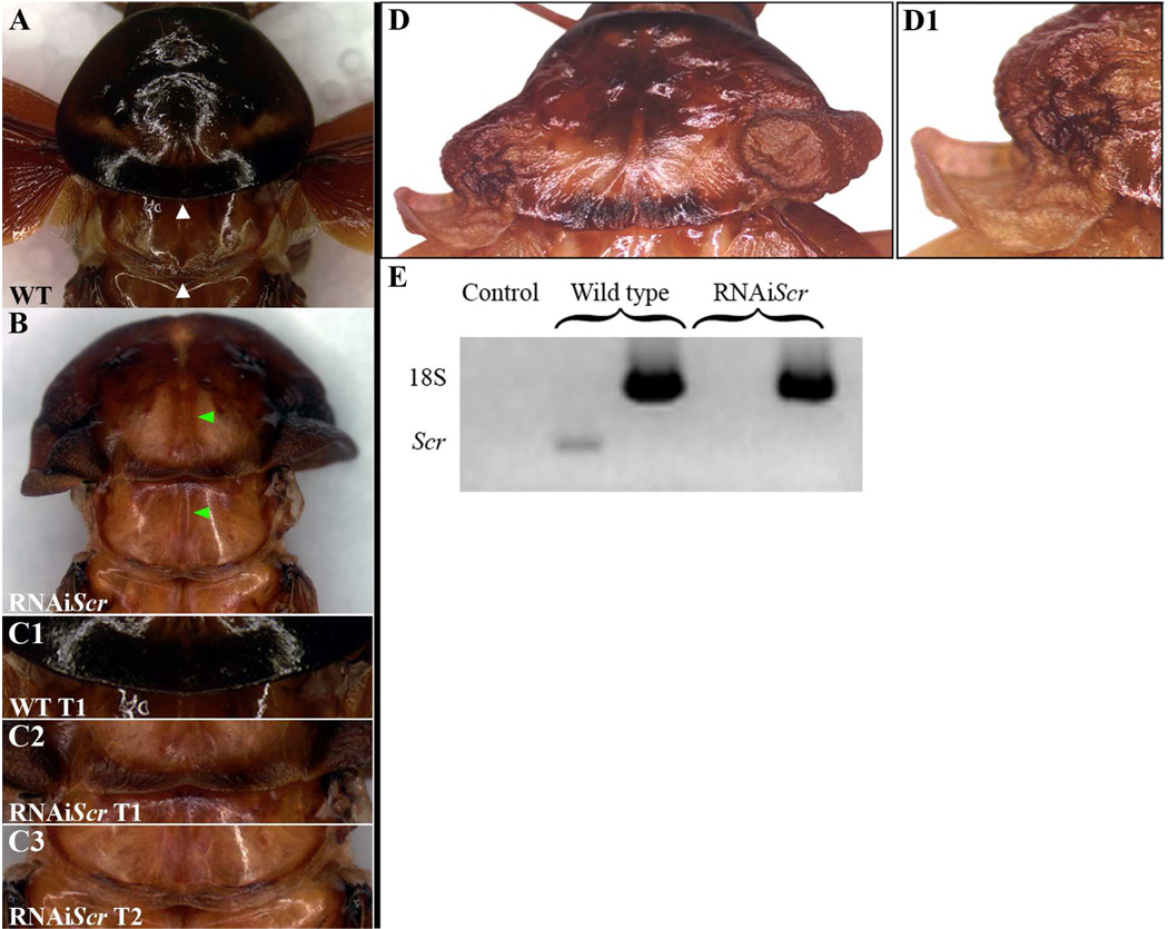
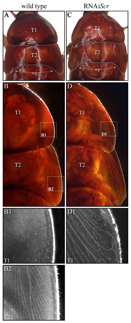
The cockroach, Periplaneta americana represents a basal insect lineage that undergoes the ancestral hemimetabolous mode of development. Here, we examine the embryonic and post-embryonic functions of the hox gene Scr in Periplaneta as a way of better understanding the roles of this gene in the evolution of insect body plans. During embryogenesis, Scr function is strictly limited to the head with no role in the prothorax. This indicates that the ancestral embryonic function of Scr was likely restricted to the head, and that the posterior expansion of expression in the T1 legs may have preceded any apparent gain of function during evolution. In addition, Scr plays a pivotal role in the formation of the dorsal ridge, a structure that separates the head and thorax in all insects. This is evidenced by the presence of a supernumerary segment that occurs between the labial and T1 segments of RNAiScr first nymphs and is attributed to an alteration in engrailed (en) expression. The fact that similar Scr phenotypes are observed in Tribolium but not in Drosophila or Oncopeltus reveals the presence of lineage-specific variation in the genetic architecture that controls the formation of the dorsal ridge. In direct contrast to the embryonic roles, Scr has no function in the head region during post-embryogenesis in Periplaneta, and instead, strictly acts to provide identity to the T1 segment. Furthermore, the strongest Periplaneta RNAiScr phenotypes develop ectopic wing-like tissue that originates from the posterior region of the prothoracic segment. This finding provides a novel insight into the current debate on the morphological origin of insect wings.
Copyright 2010 Elsevier Inc. All rights reserved.
Figures
Similar articles
-
Diverging functions of Scr between embryonic and post-embryonic development in a hemimetabolous insect, Oncopeltus fasciatus.Dev Biol. 2009 May 1;329(1):142-51. doi: 10.1016/j.ydbio.2009.01.032. Dev Biol. 2009. PMID: 19382295 Free PMC article.
-
Evolving expression patterns of the homeotic gene Scr in insects.Int J Dev Biol. 2010;54(5):897-904. doi: 10.1387/ijdb.082839kp. Int J Dev Biol. 2010. PMID: 20336613
-
The contribution of the melanin pathway to overall body pigmentation during ontogenesis of Periplaneta americana.Insect Sci. 2016 Aug;23(4):513-9. doi: 10.1111/1744-7917.12356. Epub 2016 Jun 30. Insect Sci. 2016. PMID: 27158782
-
Evolution of nubbin function in hemimetabolous and holometabolous insect appendages.Dev Biol. 2011 Sep 1;357(1):83-95. doi: 10.1016/j.ydbio.2011.06.014. Epub 2011 Jun 25. Dev Biol. 2011. PMID: 21708143 Free PMC article.
-
Proboscipedia represses distal signaling in the embryonic gnathal limb fields of Tribolium castaneum.Dev Genes Evol. 2003 Mar;213(2):55-64. doi: 10.1007/s00427-002-0291-7. Epub 2003 Jan 21. Dev Genes Evol. 2003. PMID: 12632174
Cited by
-
Upregulation of Hox genes leading to caste-specific morphogenesis in a termite.Evodevo. 2023 Jul 27;14(1):12. doi: 10.1186/s13227-023-00216-w. Evodevo. 2023. PMID: 37501210 Free PMC article.
-
Establishment of molecular genetic approaches to study gene expression and function in an invasive hemipteran, Halyomorpha halys.Evodevo. 2017 Oct 18;8:15. doi: 10.1186/s13227-017-0078-6. eCollection 2017. Evodevo. 2017. PMID: 29075432 Free PMC article.
-
Rhinoceros beetle horn development reveals deep parallels with dung beetles.PLoS Genet. 2018 Oct 4;14(10):e1007651. doi: 10.1371/journal.pgen.1007651. eCollection 2018 Oct. PLoS Genet. 2018. PMID: 30286074 Free PMC article.
-
Tergal and pleural structures contribute to the formation of ectopic prothoracic wings in cockroaches.R Soc Open Sci. 2016 Aug 3;3(8):160347. doi: 10.1098/rsos.160347. eCollection 2016 Aug. R Soc Open Sci. 2016. PMID: 27853616 Free PMC article.
-
The genetic basis of rapidly evolving male genital morphology in Drosophila.Genetics. 2011 Sep;189(1):357-74. doi: 10.1534/genetics.111.130815. Epub 2011 Jul 12. Genetics. 2011. PMID: 21750260 Free PMC article.
References
-
- Abzhanov A, Kaufman TC. Novel regulation of the homeotic gene Scr associated with a crustacean leg-to-maxilliped appendage transformation. Development. 1999;126:1121–1128. - PubMed
-
- Angelini DR, Kaufman TC. Comparative developmental genetics and the evolution of arthropod body plans. Annu Rev Genet. 2005a;39:95–119. - PubMed
-
- Angelini DR, Kaufman TC. Insect appendages and comparative ontogenetics. Dev Biol. 2005b;286:57–77. - PubMed
-
- Angelini DR, Liu PZ, Hughes CL, Kaufman TC. Hox gene function and interaction in the milkweed bug Oncopeltus fasciatus (Hemiptera) Dev Biol. 2005;287:440–455. - PubMed
-
- Aplin AC, Kaufman TC. Homeotic transformation of legs to mouthparts by proboscipedia expression in Drosophila imaginal discs. Mech Dev. 1997;62:51–60. - PubMed
Publication types
MeSH terms
Substances
Grants and funding
LinkOut - more resources
Full Text Sources
Research Materials
