Review
. 2010 May;20(5):299-309.
doi: 10.1016/j.tcb.2010.01.009. Epub 2010 Feb 19.

The p53 orchestra: Mdm2 and Mdmx set the tone

Affiliations
Review

The p53 orchestra: Mdm2 and Mdmx set the tone

Mark Wade et al. Trends Cell Biol. 2010 May.

Abstract

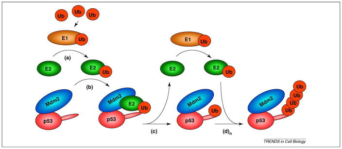
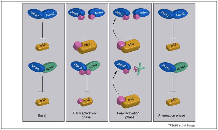
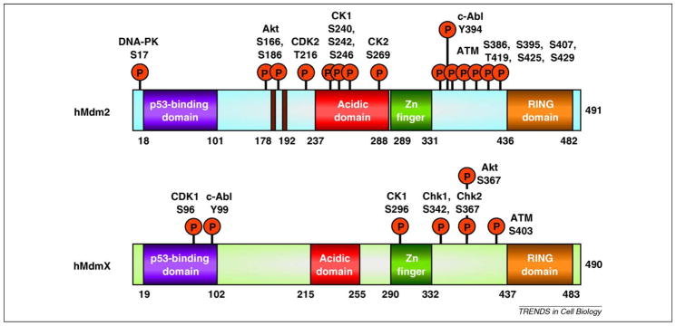
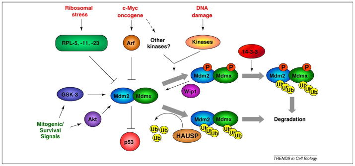
The activities of p53 cover diverse aspects of cell biology, including cell cycle control, apoptosis, metabolism, fertility, differentiation and cellular reprogramming. Although loss of p53 function engenders tumor susceptibility, hyperactivation of p53 is lethal. Therefore, p53 activity must be strictly regulated to maintain normal tissue homeostasis. Critical for the control of p53 function are its two main negative regulators: Mdm2 and Mdmx. Recent reports have provided insight into the complex mechanisms that regulate these two proteins and have revealed novel functions for each. Here, we review and evaluate models of Mdm2- and Mdmx-dependent regulation of p53 activity. Both Mdm2 and Mdmx receive input from numerous signaling pathways and interact with many proteins in addition to p53. Therefore, we also consider roles for Mdm2 and Mdmx in additional cancer-related networks, including Notch signaling and the epithelial-to-mesenchymal transition.


Figures

Figure 1
Figure 1
The E3 ubiquitin ligase function of Mdm2. Free ubiquitin (Ub) is ‘activated’ upon covalent binding to E1 ubiquitin activating enzyme. (a) Ubiquitin is then transferred to the E2 conjugating enzyme active site via a thioester linkage. (b) Mdm2 recruits ubiquitin-loaded E2, bringing it into proximity with p53. (c) Ubiquitin is transferred to p53, and E2 can re-enter the cycle with Ub-charged E1. (d) Ubiquitin-loaded E2 can again be recruited by Mdm2 for (n) cycles, thus extending the ubiquitin chain (n) times. Although mono ubiquitination and polyubiquitination can be performed by the same E2 in vitro, it is unclear whether this is also the case in vivo.
Figure 2
Figure 2
Control of p53 stability by Mdm2 homo-oligomers and Mdm2–Mdmx hetero-oligomers. Basal p53 levels are regulated by both Mdm2 homo-oligomers and Mdm2/Mdmx hetero-oligomers. In vitro data suggests the hetero-oligomer is a more effective p53 ligase in the absence of stress, and that Mdmx contributes significantly to p53 basal activity, but evidence for the regulatory importance of hetero-oligomers is limited. Immediately following DNA damage, during the early activation phase, Mdm2 is destabilized, and phosphorylated at residues outside the RING domain, destabilizing Mdm2 oligomers, leading to increased p53 levels. At the peak activation phase, Mdm2 degrades itself and Mdmx, which removes Mdm2–Mdm2 and Mdm2–Mdmx oligomers, leading to maximal p53 accumulation. During the activation phase, p53 also transactivates the Mdm2 gene (dashed arrow). The attenuation phase begins when DNA damage signaling abates. Kinase inhibition and phosphatase activation removes the pool of phosphorylated Mdm2 and Mdmx, leading to their stabilization. As a result, the homo- and hetero-oligomers regain p53 ubiquitin ligase activity, reducing p53 to basal levels. Although Mdmx degradation is clearly Mdm2-dependent and therefore requires hetero-oligomerization, the existence and ubiquitin ligase activity of phosphorylated Mdm2–Mdmx hetero-oligomers (early activation phase) is curently speculative. In addition to regulation of Mdm2 and Mdmx, phosphorylation of p53 during the damage response also contributes to p53 activation by decreasing the affinity for negative regulators, and increasing the affinity for transcriptional co-factors.
Figure 3
Figure 3
Phosphorylation of Mdm2 and Mdmx by cellular kinases. Both Mdm2 and Mdmx are extensively phosphorylated by kinases of different classes. These include damage-induced kinases ATM, Chk1, Chk2, DNA-PK and c-Abl, and proliferation/survival kinases including Akt, CK-1 and -2, CDK-1 and -2.
Figure 4
Figure 4
Multiple cell signaling cascades converge on Mdm2 and Mdmx. p53 activating stresses (red print) can signal through damage-dependent and independent pathways to activate p53. Ribosomal stress proceeds via the release of ribosomal proteins that inhibit Mdm2 ubiquitin ligase activity and stabilize p53. Oncogenes such as c-Myc can engage damage-independent pathways such as the Arf tumor suppressor in order to activate p53, or may induce DNA damage and other kinases, which then phosphorylate either Mdm2 or Mdmx. Following genotoxic stress, multiple damage-activated kinases phosphorylate Mdm2 and Mdmx and inhibit their ligase activity. This can be via increased ubiquitylation and degradation of Mdm2, and increased ubiquitylation and degradation of Mdmx, which requires binding to 14-3-3 proteins. Following resolution of a DNA damage response, the Wip1 phosphatase can dephosphorylate Mdm2 and Mdmx, leading to their stabilization. In non-stressed cells, the levels of Mdm2 and Mdmx ubiquitylation are in part controlled by the deubiquitylase HAUSP, which removes ubiquitin from each protein, leading to their stabilization. Kinases associated with proliferation and survival (green print) can also phosphorylate Mdm2 and enhance its p53 inhibitory function.

Similar articles

Cited by

References

    1. Marine JC, et al. Keeping p53 in check: essential and synergistic functions of Mdm2 and Mdm4. Cell Death Differ. 2006;13:927–934. - PubMed
    1. Kruse JP, Gu W. Modes of p53 regulation. Cell. 2009;137:609–622. - PMC - PubMed
    1. Wang YV, et al. Quantitative analyses reveal the importance of regulated Hdmx degradation for p53 activation. Proc Natl Acad Sci U S A. 2007;104:12365–12370. - PMC - PubMed
    1. Ohtsubo C, et al. Cytoplasmic tethering is involved in synergistic inhibition of p53 by Mdmx and Mdm2. Cancer Sci. 2009;100:1291–1299. - PMC - PubMed
    1. Moll UM, Petrenko O. The MDM2-p53 interaction. Mol Cancer Res. 2003;1:1001–1008. - PubMed

Publication types

MeSH terms