. 2010 Apr 16;285(16):12279-88.
doi: 10.1074/jbc.M109.086405. Epub 2010 Feb 19.

Calcium activates Nedd4 E3 ubiquitin ligases by releasing the C2 domain-mediated auto-inhibition

Affiliations

Calcium activates Nedd4 E3 ubiquitin ligases by releasing the C2 domain-mediated auto-inhibition

Jian Wang et al. J Biol Chem. .

Abstract

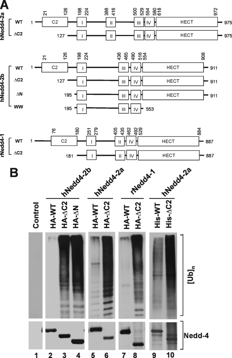
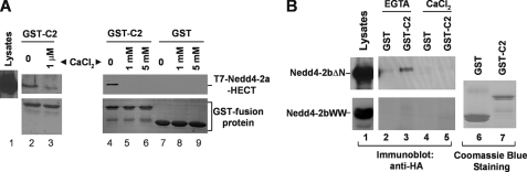
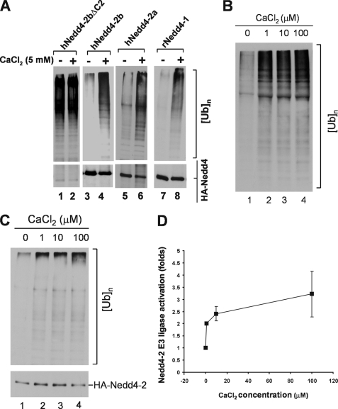
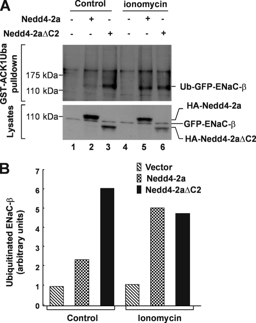
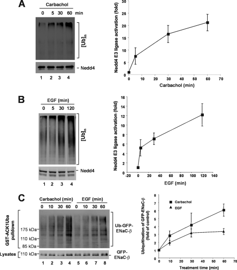
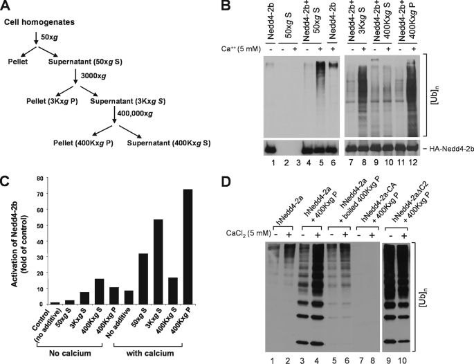
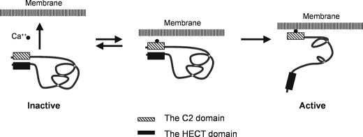
Nedd4 E3 ligases are members of the HECT E3 ubiquitin ligase family and regulate ubiquitination-mediated protein degradation. In this report, we demonstrate that calcium releases the C2 domain-mediated auto-inhibition in both Nedd4-1 and Nedd4-2. Calcium disrupts binding of the C2 domain to the HECT domain. Consistent with this, calcium activates the E3 ubiquitin ligase activity of Nedd4. Elevation of intracellular calcium by ionomycin treatment, or activation of acetylcholine receptor or epidermal growth factor receptor by carbachol or epidermal growth factor stimulation induced activation of endogenous Nedd4 in vivo evaluated by assays of either Nedd4 E3 ligase activity or ubiquitination of Nedd4 substrate ENaC-beta. The activation effect of calcium on Nedd4 E3 ligase activity was dramatically enhanced by a membrane-rich fraction, suggesting that calcium-mediated membrane translocation through the C2 domain might be an activation mechanism of Nedd4 in vivo. Our studies have revealed an activation mechanism of Nedd4 E3 ubiquitin ligases and established a connection of intracellular calcium signaling to regulation of protein ubiquitination.


Figures

FIGURE 1.
FIGURE 1.
Truncation of the C2 domain activates the E3 ubiquitin ligase activity of Nedd4. A, schematic representation of the C2 domain deletion mutants of Nedd4-1 and Nedd4-2. The roman numbers I–IV refer to the WW domains 1 (WW1), 2 (WW2), 3 (WW3), and 4 (WW4). Human Nedd4-2a and Nedd4-2b (hNedd4-2a and hNedd4-2b) are Nedd4-2 splicing forms. The Nedd4-2b lacks the WW2 domain in Nedd4-2a. Rat Nedd4-1 (rNedd4-1) has only three WW domains. The positions for the deletions and the boundaries of the domains are labeled with the amino acid residue position numbers in the peptides. B, truncation of the C2 domain activates Nedd4. Lanes 1–8, HA-tagged wild-type or the C2 domain deletion mutant of Nedd4-1 and Nedd4-2 was transfected into HEK293 cells. Nedd4 protein was immunoprecipitated with anti-HA antibody and used for the E3 ubiquitin ligase activity assay. Lanes 9 and 10, His-tagged Nedd4-2a or its C2 domain deletion mutant was expressed in E. coli and purified with affinity beads. Purified His-tagged Nedd4 and the mutant (100 ng) were used for the ubiquitin ligase activity assay. In the assay, mono-ubiquitin was used as the substrate. The ubiquitin ligation products (poly-ubiquitins) were detected by immunoblotting with anti-ubiquitin antibody. Top panels, poly-ubiquitin products; bottom panels, HA- or his-tagged wild-type and deletion mutants of Nedd4s.
FIGURE 2.
FIGURE 2.
The C2 domain of Nedd4 interacts with its HECT domain in the absence of calcium. GST-C2 domain pulldown assay for binding of the C2 domain to purified T7-tagged HECT domain of Nedd4-2a (A), or HA-tagged Nedd4-2bΔN (B). The C2 domain of Nedd4-2 was subcloned into a GST fusion protein expression vector, expressed in E. coli, and purified by affinity purification with glutathione-conjugated agarose beads. The beads with GST (control) or GST-Nedd4-2b C2 domain fusion protein were incubated with purified T7-tagged HECT domain of Nedd4-2a in indicated calcium concentrations, or with HA-Nedd4-2bΔN- or HA-Nedd4-2bWW-expressed HEK293 cell lysates in the presence of 5 mm CaCl2 or 1 mm EGTA. The GST-C2 domain-precipitated T7-Nedd4-2a-HECT, HA-Nedd4-2bΔN, or HA-Nedd4-2bWW was detected by immunoblotting with anti-T7 antibody or anti-HA antibody. The GST and GST fusion proteins used for the pulldown assay are shown by Coomassie Blue staining. Lane 1 in A and B shows one-tenth the total input of the indicated proteins in the pulldown assay.
FIGURE 3.
FIGURE 3.
Calcium activates Nedd4 E3 ubiquitin ligase activity. A, HA-tagged human Nedd4-2a, Nedd4-2b, Nedd4-2bΔC2, or rat Nedd4-1 was transfected into HEK293 cells, immunoprecipitated from the cell lysates, and used for the E3 ubiquitin ligase activity assay with or without addition of 5 mm CaCl2. The polyubiquitin products were detected by immunoblotting with anti-ubiquitin antibody (top panels), and the Nedd4 protein amount used in the ligase assay was detected by immunoblotting with anti-HA antibody (bottom panels). B, calcium activation assayed with purified Nedd4-2. His-tagged Nedd4-2a was expressed in E. coli and purified with affinity beads. Purified his-tagged Nedd4-2a (100 ng) was used for the ubiquitin ligase activity assay in the presence of indicated concentration of CaCl2. C and D, calcium activates immunoprecipitated Nedd4 E3 ubiquitin ligase from HEK293 cells. HA-tagged Nedd4-2a was transfected into HEK293 cells and immunoprecipitated with anti-HA antibody from cell homogenates. The E3 ubiquitin ligase activity of Nedd4-2a was assayed in presence of CaCl2 at indicated concentrations. C, a representative immunoblotting data; D, quantification of immunoblotting data from two independent assays of calcium-induced activation of Nedd4-2a E3 ubiquitin ligase activity by Kodak EDAS290 image system. The activation fold is defined as ([UB]n/[HA-Nedd4-2a])/([[UB]n]0/[HA-Nedd4-2a]0), where [UB]n stands for the quantity of poly-ubiquitin products at any concentration of calcium and [HA-Nedd4-2a] for the quantity of the corresponding HA-Nedd4-2a used in the assay; [[UB]n]0 for the quantity of poly-ubiquitin products with no calcium and [HA-Nedd4-2a]0 for the quantity of HA-Nedd4-2a used in the assay with no calcium.
FIGURE 4.
FIGURE 4.
Influx of extracellular calcium by ionomycin enhances ubiquitination of ENaC-β subunit, a specific Nedd4 substrate. A, a stably GFP-ENaC-β-expressed HEK293 cells transfected 36 h with HA-Nedd4-2a, Nedd4-2aΔC2, or vector were serum-starved for 12 h followed by incubation in calcium buffer (20 mm Hepes, pH 7.4, 140 mm NaCl, 6 mm KCl, 1 mm MgCl2, 1.25 mm CaCl2, 0.1 mm EDTA, and 20 mm glucose) along with or without 1 μm ionomycin for 6 h in presence of 10 μm MG-132, a proteasome inhibitor. The ubiquitinated GFP-ENaC-β was precipitated by GST-ACK1-Uba and detected by immunoblotting with anti-GFP antibody (top panel). The expression level of GFP-ENaC-β, HA-Nedd4-2a, or HA-Nedd4-2aΔC2 was determined by immunoblotting of the cell lysates with anti-GFP and anti-HA, respectively (bottom panel). B, the quantification of ubiquitinated GFP-ENaC-β in (A) with GE-Gel Logic100 Imaging system.
FIGURE 5.
FIGURE 5.
In vivo activation of Nedd4 E3 ubiquitin ligase activity by stimulation of acetylcholine receptor and EGFR. A, HEK293 cells were starved in serum-free Dulbecco's modified Eagle's medium for 2 h, then stimulated with 5 mm carbachol at the indicated time. B, human breast cancer cell MDA-MB-231 cells were serum-starved overnight followed by stimulation with EGF (100 ng/ml) at indicated time. In both A and B, endogenous Nedd4 was immunoprecipitated with an anti-Nedd4 antibody from 20,000 × g cleared cell homogenates. The E3 ubiquitin ligase activity was assayed. The poly-ubiquitin products were determined by immunoblotting and quantified by using a Kodak EDAS290 imaging system for two independent experiments. C, stimulation of acetylcholine receptor by carbachol or EGFR by EGF enhances ubiquitination of Nedd4 substrate ENaC-β subunit. GFP-ENaC-β-expressed HEK293 cells were serum-starved for 12 h, then stimulated with 5 μm carbachol or 100 ng/ml EGF at the indicated times in the presence of 10 μm MG-132. The ubiquitinated GFP-ENaC-β was precipitated by GST-ACK1-Uba and detected by immunoblotting with anti-GFP (top panel). The expression level of GFP-ENaC-β was determined by immunoblotting of the cell lysates with anti-GFP (bottom panel). Ubiquitinated GFP-ENaC-β was quantified with normalized GFP-ENaC-β by using a GE-Gel Logic100 imaging system for two independent experiments.
FIGURE 6.
FIGURE 6.
One or more heat-sensitive components enhance calcium-induced activation of Nedd4 E3 ubiquitin ligases. A, schematic procedures for preparation of cell homogenate fractions by centrifugation. B, the HA-tagged Nedd4-2b was transfected in HEK293 cells and immunoprecipitated with an anti-HA antibody for the E3-ubiquitin ligase activity assay. The preparation of the homogenate fractions of HEK293 cells and the assay of E3 ligase activity were performed as described under “Experimental Procedures.” To examine the effect of the homogenate fractions on the E3 ubiquitin ligase activity of HA-Nedd4-2b, 5 μl of the homogenate fraction was added to the reaction mix during the ubiquitin ligase activity assay. The top panels represent poly-ubiquitin products generated from the E3 ubiquitin ligase assay; the bottom panels show amounts of immunoprecipitated HA-tagged Nedd4-2b used in the assay. S50, the 50 × g supernatant; S3K, the 3,000 × g supernatant; P3K, the 3,000 × g pellet; S96K, the 96,000 × g supernatant; P96K, the 96,000 × g pellet; S400K, 400,000 × g supernatant; and P400K, the 400,000 × g pellet. C, the poly-ubiquitin product in B was quantified by using the GE-Gel Logic100 Imaging system and used for calculation of Nedd4-2b activation. D, purified Nedd4-2a is activated by the component in the 400,000 × g pellet. His-tagged Nedd4-2a was expressed in E. coli and purified with affinity beads. Purified His-tagged Nedd4-2a (100 ng), active mutant Nedd4-2aΔC2 (100 ng), or ligase-dead mutant Nedd4-2a-CA (100 ng) was used for the ubiquitin ligase activity assay in the presence or absence of CaCl2 with addition of 5 μl of indicated homogenate fraction. The poly-ubiquitin product was detected by immunoblotting with anti-ubiquitin antibody.
FIGURE 7.
FIGURE 7.
A model for activation of Nedd4 in cells. Cellular signaling elevates the cytoplasmic concentration of calcium, which binds to the C2 domain and enables the C2 domain to interact with the cellular membrane. Calcium-dependent binding of the C2 domain to the membrane results in dissociation of the C2 domain from the HECT domain, and release of the C2 domain-mediated auto-inhibition thus activates Nedd4 E3 ligase activity.

References

    1. Pickart C. M. (2001) Annu. Rev. Biochem. 70, 503–533 - PubMed
    1. Jackson P. K., Eldridge A. G., Freed E., Furstenthal L., Hsu J. Y., Kaiser B. K., Reimann J. D. (2000) Trends Cell Biol. 10, 429–439 - PubMed
    1. Passmore L. A., Barford D. (2004) Biochem. J. 379, 513–525 - PMC - PubMed
    1. Shearwin-Whyatt L., Dalton H. E., Foot N., Kumar S. (2006) BioEssays 28, 617–628 - PubMed
    1. Chen H., Ross C. A., Wang N., Huo Y., MacKinnon D. F., Potash J. B., Simpson S. G., McMahon F. J., DePaulo J. R., Jr., McInnis M. G. (2001) Eur. J. Hum. Genet. 9, 922–930 - PubMed

Publication types

MeSH terms