

Long-term functional improvement and gene expression changes after bone marrow-derived multipotent progenitor cell transplantation in myocardial infarction
- PMID: 20173039
- PMCID: PMC3774483
- DOI: 10.1152/ajpheart.01100.2009
Long-term functional improvement and gene expression changes after bone marrow-derived multipotent progenitor cell transplantation in myocardial infarction
Abstract
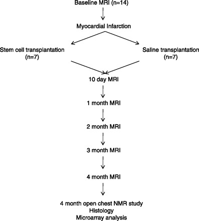
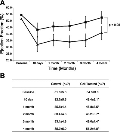
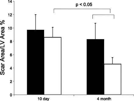
The study examined the long-term outcome of cardiac stem cell transplantation in hearts with postinfarction left ventricular (LV) remodeling. Myocardial infarction (MI) was created by ligating the first and second diagonal branches of the left anterior descending coronary artery in miniature swine. Intramyocardial injections of 50 million LacZ-labeled bone marrow-derived multipotent progenitor cells (MPC) were performed in the periscar region (Cell, n = 7) immediately after MI, whereas, in control animals (Cont, n = 7), saline was injected. Functional outcome was assessed monthly for 4 mo with MRI and (31)P-magnetic resonance spectroscopy. Engraftment was studied on histology, and gene chip (Affymetrix) array analysis was used to study differential expression of genes in the two groups. MPC treatment resulted in improvement of ejection fraction as early as 10 days after MI (Cell, 43.4 +/- 5.1% vs. Cont, 32.2 +/- 5.5%; P < 0.05). This improvement was seen each month and persisted to 4 mo (Cell, 51.2 +/- 4.8% vs. Cont, 35.7 +/- 5.0%; P < 0.05). PCr-to-ATP ratio (PCr/ATP) improved with MPC transplantation, which was most pronounced at high cardiac work states (subendocardial PCr/ATP was 1.70 +/- 0.10 vs. 1.34 +/- 0.14, P < 0.05). There was no significant difference in scar size (scar/LV area * 100) at 10 days postinfarction. However, at 4 mo, there was a significant decrease in scar size in the Cell group (Cell, 4.6 +/- 1.0% vs. Cont, 8.6 +/- 2.4%; P < 0.05). No significant engraftment of MPC was observed. MPC transplantation was associated with a downregulation of mitochondrial oxidative enzymes and increased levels of myocyte enhancer factor 2a and zinc finger protein 91. In conclusion, MPC transplantation leads to long-term functional and bioenergetic improvement in a porcine model of postinfarction LV remodeling, despite no significant engraftment of stem cells in the heart. MPC transplantation reduces regional wall stresses and infarct size and mitigates the adverse effects of LV remodeling, as seen by a reduction in LV hypertrophy and LV dilatation, and is associated with differential expression of genes relating to metabolism and apoptosis.
Figures

Comment in
-
Long-term effects of stem cell transplantation in the postinfarct heart: benefits and mechanisms.Am J Physiol Heart Circ Physiol. 2010 May;298(5):H1308-9. doi: 10.1152/ajpheart.00192.2010. Epub 2010 Mar 5. Am J Physiol Heart Circ Physiol. 2010. PMID: 20207819 No abstract available.
Similar articles
-
Bioenergetic and functional consequences of bone marrow-derived multipotent progenitor cell transplantation in hearts with postinfarction left ventricular remodeling.Circulation. 2007 Apr 10;115(14):1866-75. doi: 10.1161/CIRCULATIONAHA.106.659730. Epub 2007 Mar 26. Circulation. 2007. PMID: 17389266
-
Stem cells for myocardial repair with use of a transarterial catheter.Circulation. 2009 Sep 15;120(11 Suppl):S238-46. doi: 10.1161/CIRCULATIONAHA.109.885236. Circulation. 2009. PMID: 19752374 Free PMC article.
-
Relationships between regional myocardial wall stress and bioenergetics in hearts with left ventricular hypertrophy.Am J Physiol Heart Circ Physiol. 2008 May;294(5):H2313-21. doi: 10.1152/ajpheart.01288.2007. Epub 2008 Mar 7. Am J Physiol Heart Circ Physiol. 2008. PMID: 18326803 Free PMC article.
-
Cellular therapy promotes endogenous stem cell repair.Can J Physiol Pharmacol. 2012 Oct;90(10):1335-44. doi: 10.1139/y2012-115. Epub 2012 Sep 28. Can J Physiol Pharmacol. 2012. PMID: 23020202 Free PMC article. Review.
-
Myocardial remodeling: cellular and extracellular events and targets.Annu Rev Physiol. 2011;73:47-68. doi: 10.1146/annurev-physiol-012110-142230. Annu Rev Physiol. 2011. PMID: 21314431 Free PMC article. Review.
Cited by
-
Mesenchymal stem cell transplantation for the infarcted heart: a role in minimizing abnormalities in cardiac-specific energy metabolism.Am J Physiol Endocrinol Metab. 2012 Jan 15;302(2):E163-72. doi: 10.1152/ajpendo.00443.2011. Epub 2011 Oct 4. Am J Physiol Endocrinol Metab. 2012. PMID: 21971524 Free PMC article.
-
Molecular imaging of mesenchymal stem cell: mechanistic insight into cardiac repair after experimental myocardial infarction.Circ Cardiovasc Imaging. 2012 Jan;5(1):94-101. doi: 10.1161/CIRCIMAGING.111.966424. Epub 2011 Dec 1. Circ Cardiovasc Imaging. 2012. PMID: 22135400 Free PMC article.
-
Evidence for Mechanisms Underlying the Functional Benefits of a Myocardial Matrix Hydrogel for Post-MI Treatment.J Am Coll Cardiol. 2016 Mar 8;67(9):1074-1086. doi: 10.1016/j.jacc.2015.12.035. J Am Coll Cardiol. 2016. PMID: 26940929 Free PMC article.
-
Effects of maternal immune activation in porcine transcript isoforms of neuropeptide and receptor genes.J Integr Neurosci. 2021 Mar 30;20(1):21-31. doi: 10.31083/j.jin.2021.01.332. J Integr Neurosci. 2021. PMID: 33834688 Free PMC article.
-
The benefit of enhanced contractility in the infarct borderzone: a virtual experiment.Front Physiol. 2012 Apr 9;3:86. doi: 10.3389/fphys.2012.00086. eCollection 2012. Front Physiol. 2012. PMID: 22509168 Free PMC article.
References
-
- Amado LC, Saliaris AP, Schuleri KH, St John M, Xie JS, Cattaneo S, Durand DJ, Fitton T, Kuang JQ, Stewart G, Lehrke S, Baumgartner WW, Martin BJ, Heldman AW, Hare JM. Cardiac repair with intramyocardial injection of allogeneic mesenchymal stem cells after myocardial infarction. Proc Natl Acad Sci USA 102: 11474–11479, 2005 - PMC - PubMed
-
- Assmus B, Honold J, Schachinger V, Britten MB, Fischer-Rasokat U, Lehmann R, Teupe C, Pistorius K, Martin H, Abolmaali ND, Tonn T, Dimmeler S, Zeiher AM. Transcoronary transplantation of progenitor cells after myocardial infarction. N Engl J Med 355: 1222–1232, 2006 - PubMed
-
- Assmus B, Schachinger V, Teupe C, Britten M, Lehmann R, Dobert N, Grunwald F, Aicher A, Urbich C, Martin H, Hoelzer D, Dimmeler S, Zeiher AM. Transplantation of Progenitor Cells and Regeneration Enhancement in Acute Myocardial Infarction (TOPCARE-AMI). Circulation 106: 3009–3017, 2002 - PubMed
-
- Black BL, Olson EN. Transcriptional control of muscle development by myocyte enhancer factor-2 (MEF2) proteins. Annu Rev Cell Dev Biol 14: 167–196, 1998 - PubMed
-
- Chen W, Cho Y, Merkle H, Ye Y, Zhang Y, Gong G, Zhang J, Ugurbil K. In vitro and in vivo studies of 1H NMR visibility to detect deoxyhemoglobin and deoxymyoglobin signals in myocardium. Magn Reson Med 42: 1–5, 1999 - PubMed
Publication types
MeSH terms
Substances
Grants and funding
LinkOut - more resources
Full Text Sources
Other Literature Sources
Medical
Molecular Biology Databases