. 2010 May;298(5):H1518-28.
doi: 10.1152/ajpheart.00813.2009. Epub 2010 Feb 19.

Mice deficient in Mkp-1 develop more severe pulmonary hypertension and greater lung protein levels of arginase in response to chronic hypoxia

Affiliations

Mice deficient in Mkp-1 develop more severe pulmonary hypertension and greater lung protein levels of arginase in response to chronic hypoxia

Yi Jin et al. Am J Physiol Heart Circ Physiol. 2010 May.

Abstract

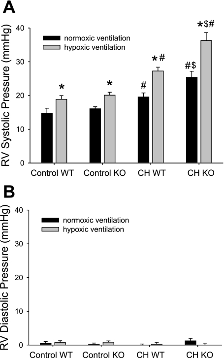
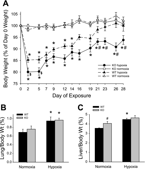
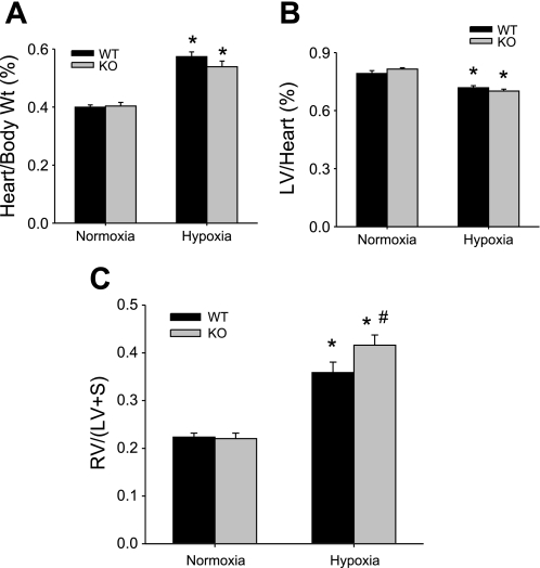
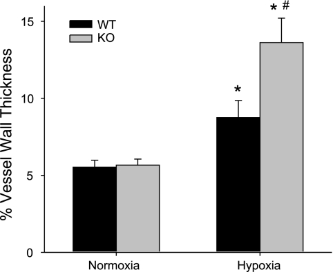
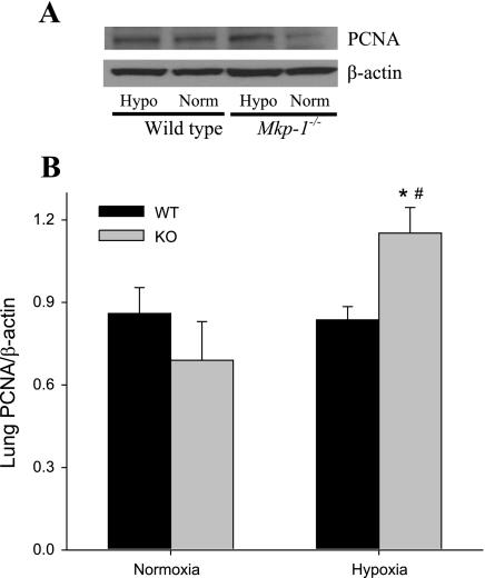
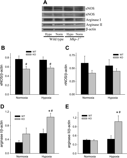
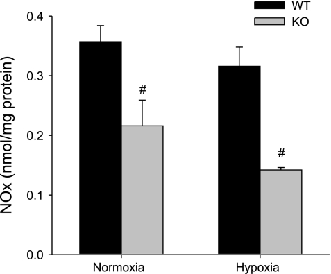
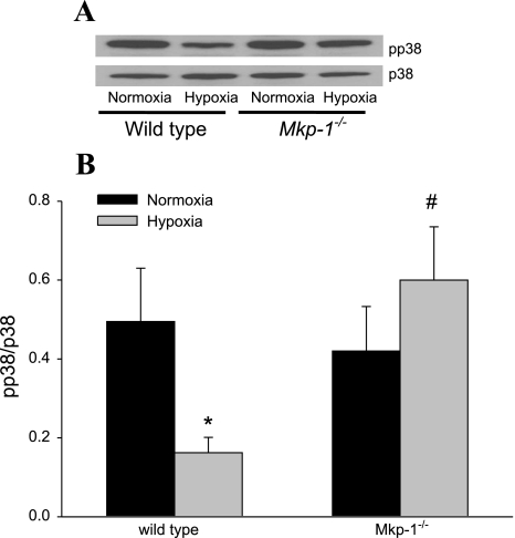
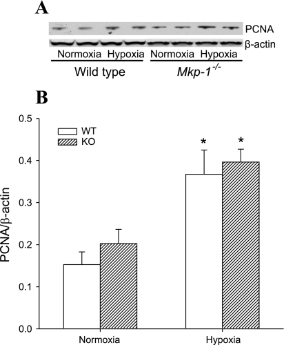
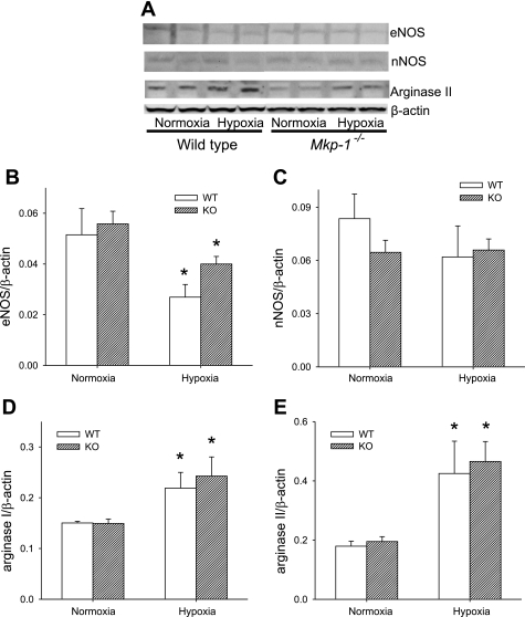
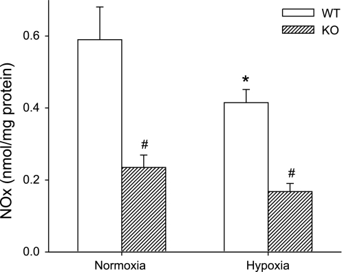
The mitogen-activated protein (MAP) kinases are involved in cellular responses to many stimuli, including hypoxia. MAP kinase signaling is regulated by a family of phosphatases that include MAP kinase phosphatase-1 (MKP-1). We hypothesized that mice lacking the Mkp-1 gene would have exaggerated chronic hypoxia-induced pulmonary hypertension. Wild-type (WT) and Mkp-1(-/-) mice were exposed to either 4 wk of normoxia or hypobaric hypoxia. Following chronic hypoxia, both genotypes demonstrated elevated right ventricular pressures, right ventricular hypertrophy as demonstrated by the ratio of the right ventricle to the left ventricle plus septum weights [RV(LV + S)], and greater vascular remodeling. However, the right ventricular systolic pressures, the RV/(LV + S), and the medial wall thickness of 100- to 300-microm vessels was significantly greater in the Mkp-1(-/-) mice than in the WT mice following 4 wk of hypobaric hypoxia. Chronic hypoxic exposure caused no detectable change in eNOS protein levels in the lungs in either genotype; however, Mkp-1(-/-) mice had lower levels of eNOS protein and lower lung NO production than did WT mice. No iNOS protein was detected in the lungs by Western blotting in any condition in either genotype. Both arginase I and arginase II protein levels were greater in the lungs of hypoxic Mkp-1(-/-) mice than those in hypoxic WT mice. Lung levels of proliferating cell nuclear antigen were greater in hypoxic Mkp-1(-/-) than in hypoxic WT mice. These data are consistent with the concept that MKP-1 acts to restrain hypoxia-induced arginase expression and thereby reduces vascular remodeling and the severity of pulmonary hypertension.


Figures

Fig. 1.
Fig. 1.
Chronic hypoxia (CH) results in higher right ventricular (RV) pressures, and mitogen-activated protein (MAP) kinase phosphatase-1 (MKP)-1-deficient animals had higher RV pressures after chronic hypoxia than did wild-type (WT) animals. A: RV systolic pressures (RVSP) during 5 min of normoxia (black bars, FiO2 0.21), followed by 10 min of hypoxic (gray bars, FiO2 0.12), ventilation in anesthetized mice. Control animals were exposed to room air, whereas CH animals were exposed to hypobaric hypoxia (barometric pressure ≈ 380 mmHg) for 4 wk before study. Both WT and Mkp-1−/− [knockout (KO)] mice were studied. There were 5 animals in each study group, except for the CH KO, which had 4 animals. P < 0.05, hypoxic ventilation different from normoxic ventilation in the anesthetized animals in the same group (*), chronic hypoxia-exposed animals different from control animals in the same genotype and ventilation (#), and CH KO animals different from CH WT animals in the same ventilation condition ($). B: RV diastolic pressures from the same animals as in A. There were no differences in diastolic pressures with either ventilation, exposure, or genotype.
Fig. 2.
Fig. 2.
MKP-1 deficiency results in sustained weight loss during chronic hypoxic exposure. A: body weight change (as a percent of the day 0 weight) during exposure in normoxic WT mice (▵), hypoxic WT mice (▲), normoxic Mkp-1−/− (KO) mice (○), and hypoxic Mkp-1−/− mice (●). Both genotypes lost weight initially during hypoxic exposure, but only Mkp-1−/− mice had significant weight loss by the end of the exposure period. P < 0.05, hypoxia different from normoxia same genotype (*), hypoxic Mkp-1−/− mice different from hypoxic WT mice ($), and hypoxic Mkp-1−/− mice different from 3 other study groups (#); n = 6 mice in each group. B: the ratio of lung-to-body weight in WT mice (black bars) or in Mkp-1−/− mice (gray bars, n = 6 in each group). Hypoxic exposure resulted in greater lung weights in both genotypes. *P < 0.05, hypoxia different from normoxia same genotype. C: the ratio of liver-to-body weight in WT mice (black bars) or in Mkp-1−/− mice (gray bars, n = 6 in each group). Liver weight was only increased in WT in hypoxia. P < 0.05, hypoxia different from normoxia same genotype (*) and Mkp-1−/− different from WT same condition (#).
Fig. 3.
Fig. 3.
Chronic hypoxia-induced right ventricular hypertrophy was greater in Mkp-1−/− mice than in WT mice. A: the ratio of total heart-to-body weight in WT mice (black bars) and in Mkp-1−/− mice (gray bars, n = 6 in each group). Chronic hypoxia resulted in greater total heart weight in both genotypes. *P < 0.05, hypoxia different from normoxia same genotype. B: the ratio of left ventricular-to-total heart weight in WT mice (black bars) or in Mkp-1−/− mice (gray bars, n = 6 in each group). Chronic hypoxia resulted in lower left ventricle (LV) weights in both genotypes. *P < 0.05, hypoxia different from normoxia same genotype. C: the ratio of RV/[LV + septum (S)] in WT mice (black bars) or in Mkp-1−/− mice (gray bars, n = 6 in each group). RV/(LV + S) was increased by chronic hypoxia and was greater in the Mkp-1−/− mice than in the WT mice. P < 0.05, hypoxia different from normoxia same genotype (*) and Mkp-1−/− different from WT same condition (#).
Fig. 4.
Fig. 4.
Chronic hypoxia exposure resulted in greater vessel wall thickness in the lungs from Mkp-1−/− mice than in vessels from the lungs of WT mice. The percent vessel wall thickness from pulmonary arteries of 100 to 300 μm outer diameter from WT and Mkp-1−/− mice in normoxia and hypoxia. Hypoxia resulted in significant vascular remodeling in both genotypes; however, there was significantly greater wall thickness in the chronically hypoxic Mkp-1−/− mice than in the chronically hypoxic WT mice. The vessels were taken from lung sections from 3 animals in each group; the number of vessels is 31 for WT normoxia, 9 for Mkp-1−/− normoxia, 33 for WT hypoxia, and 43 for Mkp-1−/− hypoxia. P < 0.05, hypoxia different from normoxia same genotype (*) and Mkp-1−/− different from WT same condition (#).
Fig. 5.
Fig. 5.
Deficiency in MKP-1 resulted in higher proliferating cell nuclear antigen (PCNA) protein expression in the lungs following chronic hypoxia exposure. A: representative Western blot for PCNA in lung tissue. Note that the hypoxic (Hypo) tissues are in lanes 1 and 3, and the normoxic (Norm) tissues are in lanes 2 and 4. B: the mean densities for PCNA protein normalized to β-actin protein in the lung of WT (black bars) and Mkp-1−/− (gray bars) mice; n = 6 for each group. P < 0.05, hypoxia different from normoxia same genotype (*) and Mkp-1−/− different from WT same condition (#).
Fig. 6.
Fig. 6.
After 4 wk of hypoxia, there was no difference in lung nitric oxide synthase (NOS) protein levels, but lung arginase levels were increased only in hypoxic Mkp-1−/− mice. A: representative Western blots for endothelial NOS (eNOS), neuronal NOS (nNOS), arginase I, arginase II, and β-actin for lung tissue from WT and Mkp-1−/− mice after 4 wk in normoxia or hypoxia. Note that the hypoxic tissues are in lanes 1 and 3, and the normoxic tissues are in lanes 2 and 4. B: the mean densities for eNOS protein normalized to β-actin protein in the lung of WT (black bars) and Mkp-1−/− (gray bars) mice. The levels of eNOS protein were significantly lower in the Mkp-1−/− mice than in the WT mice. C: the mean densities for nNOS protein normalized to β-actin protein in the lung of WT (black bars) and Mkp-1−/− (gray bars) mice. The levels of nNOS protein were not significantly different between genotypes. We were unable to detect inducible NOS (iNOS) in the lungs from any of the groups of mice studied. D: the mean densities for arginase I protein normalized to β-actin protein in the lungs of WT (black bars) and Mkp-1−/− (gray bars) mice. E: the mean densities for arginase II protein normalized to β-actin protein in the lungs of WT (black bars) and Mkp-1−/− (gray bars) mice; n = 5–6. P < 0.05, hypoxia different from normoxia same genotype (*) and Mkp-1−/− different from WT same condition (#).
Fig. 7.
Fig. 7.
Deficiency in MKP-1 decreased NO production in the lungs. Accumulation of nitrite/nitrate (NOx) in the lungs from WT (black bars) and Mkp-1−/− (gray bars) mice under normoxia or hypoxia was measured using a chemiluminescence NO analyzer and normalized to lung protein concentration (nmol/mg protein); n = 5–6 in each group. #P < 0.05, Mkp-1−/− different from WT same condition.
Fig. 8.
Fig. 8.
Deficiency of MKP-1 resulted in greater phosphorylated p38 (pp38) expression after 5 days in hypoxia. A: representative Western blot for pp38, which was then stripped and reprobed for p38. B: the mean densities of pp38 normalized to total p38 in the lung of WT and Mkp-1−/− mice after 5 days in normoxia (black bars) or hypoxia (gray bars). P < 0.05, hypoxia different from normoxia same genotype (*) and hypoxia-exposed Mkp-1−/− different from hypoxia-exposed WT (#).
Fig. 9.
Fig. 9.
Chronic hypoxia resulted in higher right ventricular PCNA protein expression at 4 wk in both WT and Mkp-1−/− mice. A: representative Western blot for PCNA in right ventricular tissue. B: the mean densities for PCNA protein normalized to β-actin protein in the right ventricles of WT (open bars) and Mkp-1−/− (cross-hatched bars) mice. *P < 0.05, hypoxia different from normoxia same genotype.
Fig. 10.
Fig. 10.
Chronic hypoxia decreased eNOS protein expression in the right ventricle and increased arginase protein expression in both genotypes. A: representative Western blots for eNOS, nNOS, arginase II, and β-actin for right ventricular tissue from WT and Mkp-1−/− mice after 4 wk in normoxia or hypoxia. B: the mean densities for eNOS protein normalized to β-actin protein in the right ventricle of WT (open bars) and Mkp-1−/− (cross-hatched bars) mice. After chronic hypoxia, the levels of eNOS protein in the lung were lower than in respective control-exposed animals for both genotypes. C: the mean densities for nNOS protein normalized to β-actin protein in the right ventricle of WT (open bars) and Mkp-1−/− (cross-hatched bars) mice. We were unable to detect iNOS in the right ventricle from any of the groups of mice studied. D: the mean densities for arginase I protein normalized to β-actin protein in the right ventricle of WT (open bars) and Mkp-1−/− (cross-hatched bars) mice. After 4 wk of chronic hypoxic exposure, the protein levels of arginase were increased in both genotypes. E: the mean densities for arginase II protein normalized to β-actin protein in the right ventricle of WT (open bars) and Mkp-1−/− (cross-hatched bars) mice. After 4 wk of chronic hypoxia, the arginase II protein levels were greater in both genotypes than in their respective normoxic controls; n = 5–6 in each group. *P < 0.05, hypoxia different from normoxia same genotype.
Fig. 11.
Fig. 11.
Deficiency in MKP-1 decreased NO production in the right ventricle, whereas chronic hypoxia decreased NO production only in WT mice. Accumulation of NOx in the right ventricles from WT (open bars) and Mkp-1−/− (cross-hatched bars) mice under normoxia or hypoxia was measured using a chemiluminescence NO analyzer and normalized to right ventricular protein concentration (nmol/mg protein); n = 5–6 in each group. P < 0.05, Mkp-1−/− different from WT same condition (#) and hypoxia different from normoxia same genotype (*).

Similar articles

Cited by

References

    1. Albina JE, Mahoney EJ, Daley JM, Wesche DE, Morris SM, Jr, Reichner JS. Macrophage arginase regulation by CCAAT/enhancer-binding protein beta. Shock 23: 168–72, 2005 - PubMed
    1. Bueno OF, De Windt LJ, Lim HW, Tymitz KM, Witt SA, Kimball TR, Molkentin JD. The dual-specificity phosphatase MKP-1 limits the cardiac hypertrophic response in vitro and in vivo. Circ Res 88: 88–96, 2001 - PubMed
    1. Buermans HPJ, Redout EM, Schiel AE, Musters RJP, Zuidwijk M, Eijk PP, van Hardeveld C, Kasanmoentalib S, Visser FC, Ylstra B, Simonides WS. Microarray analysis reveals pivotal divergent mRNA expression profiles early in the development of either compensated ventricular hypertrophy or heart failure. Physiol Genomics 21: 314–323, 2005 - PubMed
    1. Case D, Irwin D, Ivester C, Harral J, Morris K, Imamura M, Roedersheimer M, Paterson A, Carr M, Hagen M, Saavedra M, Crossno J, Young KA, Dempsey EC, Poirier F, West J, Majka S. Mice deficient in galectin-1 exhibit attenuated physiological responses to chronic hypoxia-induced pulmonary hypertension. Am J Physiol Lung Cell Mol Physiol 292: L154–L164, 2007 - PubMed
    1. Calvert TJ, Chicoine LG, Liu Y, Nelin LD. Deficiency of mitogen-activated protein kinase phosphotase-1 results in iNOS-mediated hypotension in response to low-dose endotoxin. Am J Physiol Heart Circ Physiol 294: H1621–H1629, 2008 - PubMed

Publication types

MeSH terms