. 2010 Mar 9;107(10):4561-6.
doi: 10.1073/pnas.0914495107. Epub 2010 Feb 19.

Sampling the N-terminal proteome of human blood

Affiliations

Sampling the N-terminal proteome of human blood

David Wildes et al. Proc Natl Acad Sci U S A. .

Abstract

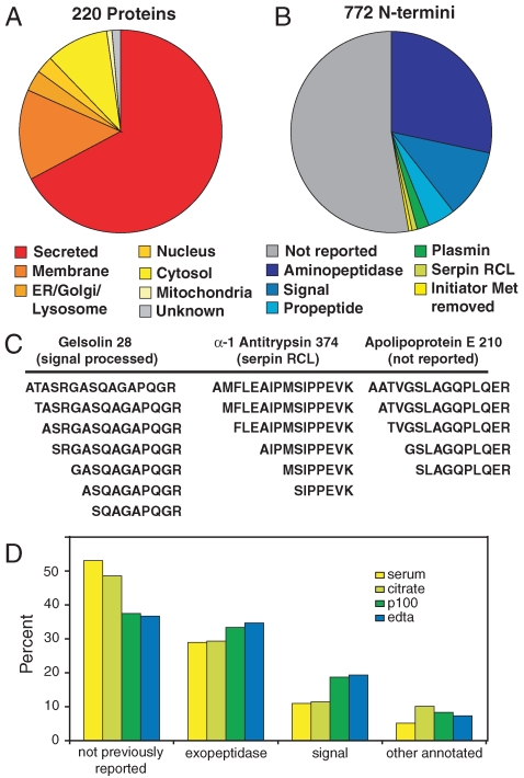
The proteomes of blood plasma and serum represent a potential gold mine of biological and diagnostic information, but challenges such as dynamic range of protein concentration have hampered efforts to unlock this resource. Here we present a method to label and isolate N-terminal peptides from human plasma and serum. This process dramatically reduces the complexity of the sample by eliminating internal peptides. We identify 772 unique N-terminal peptides in 222 proteins, ranging over six orders of magnitude in abundance. This approach is highly suited for studying natural proteolysis in plasma and serum. We find internal cleavages in plasma proteins created by endo- and exopeptidases, providing information about the activities of proteolytic enzymes in blood, which may be correlated with disease states. We also find signatures of signal peptide cleavage, coagulation and complement activation, and other known proteolytic processes, in addition to a large number of cleavages that have not been reported previously, including over 200 cleavages of blood proteins by aminopeptidases. Finally, we can identify substrates from specific proteases by exogenous addition of the protease combined with N-terminal isolation and quantitative mass spectrometry. In this way we identified proteins cleaved in human plasma by membrane-type serine protease 1, an enzyme linked to cancer progression. These studies demonstrate the utility of direct N-terminal labeling by subtiligase to identify and characterize endogenous and exogenous proteolysis in human plasma and serum.


Conflict of interest statement

The authors declare no conflict of interest.

Figures

Fig. 1.
Fig. 1.
Method for specific, enzymatic labeling of N termini in serum. (A) Schematic of workflow. Subtiligase is used to transfer a peptide containing biotin and a TEV protease-cleavable linker onto protein N termini. Proteins are then captured on streptavidin beads and trypsinized, removing all but the N-terminal tryptic peptide. Trypsinization on beads reduces unlabeled background created from sample precipitation in solution digests. N-terminal peptides are released with TEV protease for strong cation exchange fractionation and MS/MS analysis. Release leaves a SY-dipeptide tag on the N terminus.
Fig. 2.
Fig. 2.
Concentration distribution of proteins and reproducibility of the method. (A) A subset of 110 proteins of established abundance (11) is plotted by mean molar concentration in plasma. Representative low, medium, and high abundance proteins are labeled. (B) The number of N termini detected in each protein is shown, arranged in order of abundance. Proteins depicted in this plot are given in Table S1. (C) Venn diagram showing results of three replicate experiments on a single sample of citrated plasma.
Fig. 3.
Fig. 3.
The N-terminal proteome of human blood. (A) The subcellular locations, as annotated in Swiss-Prot (www.uniprot.org), of the proteins detected in this study. (B) Cleavage site annotations of detected N termini, according to Swiss-Prot and MEROPS (40) databases. (C) Evidence of aminopeptidase trimming of N termini in three proteins. Similar degradation was seen in 112 N termini. (D) Comparison of cleavage annotations found in serum and plasma collected with three different anticoagulants.
Fig. 4.
Fig. 4.
Patterns of endoproteolysis. (A) Sequence logo of the eight residues (P4–P4′) surrounding the cleavage site of all nonannotated, endoproteolytic cleavages. The y axis denotes information content and has a maximum value of 4.2. Logo created by Weblogo (http://weblogo.berkeley.edu/) (41). (C) Cleavage of two significant blood proteins, prothrombin and complement C3. Swiss-Prot annotated cleavage sites divide the proteins into domains as shown on the schematic. Cleavage sites detected in this study are indicated with arrows. AP, thrombin activation peptide; LC, thrombin light chain.
Fig. 5.
Fig. 5.
Plots of iTRAQ signal for representative putative MT-SP1 substrates. ● A2M 705; ○ A2M 707; ▴ A2M 707; ▵ A2M720; ▪ complement C3 713; □ complement C3 741.

References

    1. Anderson NL, Anderson NG. The human plasma proteome: History, character, and diagnostic prospects. Mol Cell Proteomics. 2002;1:845–867. - PubMed
    1. Liu T, et al. Evaluation of multiprotein immunoaffinity subtraction for plasma proteomics and candidate biomarker discovery using mass spectrometry. Mol Cell Proteomics. 2006;5:2167–2174. - PMC - PubMed
    1. Zolotarjova N, et al. Differences among techniques for high-abundant protein depletion. Proteomics. 2005;5:3304–3313. - PubMed
    1. Cohen MP, Clements RS. Measuring glycated proteins: Clinical and methodological aspects. Diabetes Technol The. 1999;1:57–70. - PubMed
    1. Agard NJ, Wells JA. Methods for the proteomic identification of protease substrates. Curr Opin Chem Biol. 2009;13:503–509. - PMC - PubMed

Publication types

LinkOut - more resources