Review
. 2010:11:27-60.
doi: 10.1159/000289196. Epub 2010 Feb 18.

Transcriptional control of the TNF gene

Affiliations
Review

Transcriptional control of the TNF gene

James V Falvo et al. Curr Dir Autoimmun. 2010.

Abstract

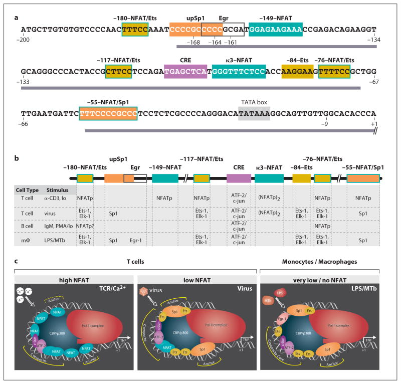
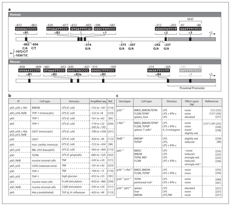
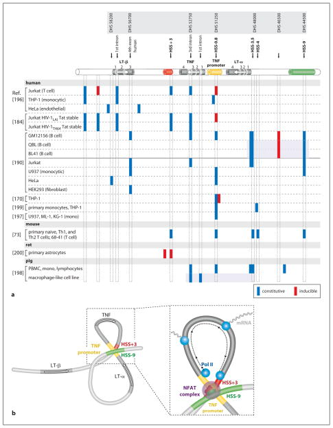
The cytokine TNF is a critical mediator of immune and inflammatory responses. The TNF gene is an immediate early gene, rapidly transcribed in a variety of cell types following exposure to a broad range of pathogens and signals of inflammation and stress. Regulation of TNF gene expression at the transcriptional level is cell type- and stimulus-specific, involving the recruitment of distinct sets of transcription factors to a compact and modular promoter region. In this review, we describe our current understanding of the mechanisms through which TNF transcription is specifically activated by a variety of extracellular stimuli in multiple cell types, including T cells, B cells, macrophages, mast cells, dendritic cells, and fibroblasts. We discuss the role of nuclear factor of activated T cells and other transcription factors and coactivators in enhanceosome formation, as well as the contradictory evidence for a role for nuclear factor kappaB as a classical activator of the TNF gene. We describe the impact of evolutionarily conserved cis-regulatory DNA motifs in the TNF locus upon TNF gene transcription, in contrast to the neutral effect of single nucleotide polymorphisms. We also assess the regulatory role of chromatin organization, epigenetic modifications, and long-range chromosomal interactions at the TNF locus.


Figures

Fig. 1
Fig. 1
Cell type- and stimulus-specific enhanceosome formation at the proximal TNF promoter. a Sequence of human proximal TNF promoter showing the positions of transcription factor binding sites (boxes), regions of highest sequence conservation in the primate lineage (bar) [33], and positions of fixed genetic differences in nonhuman primates (−9 G/T) and in orangutan and gibbon species (−168 G/A, −164 C/T, −161 C/T) [32, 33]. Adapted from Baena et al. [33]. b Transcription factors that bind and function at sites in the indicated cell types in response to the indicated stimuli [, –24, 34, 35]. c Model of the role of ambient NFAT levels upon TNF enhanceosome formation, with proteins bound at the CRE/κ3/Ets composite site interacting with ‘anchor’ complexes to stabilize interactions with CBP/p300 and the RNA Pol II complex [23, 24]. Adapted from Barthel et al. [24].
Fig. 2
Fig. 2
NF-κB and TNF gene regulation. a Putative NF-κB and NF-κB-like sites in the human and murine TNF promoters, along with the positions of SNPs in the human TNF promoter. b Stimulus-dependent association of NF-κB proteins (in at least one condition or time point studied) with the TNF promoter in ChIP assays. c Impact of deletions of NF-κB proteins in mice upon TNF gene transcription (asterisk denotes studies and cell types where only protein expression was assayed) in response to various stimuli). TEPM = Thioglycollate-elicited peritoneal macrophages; FLDM = fetal liver-derived macrophages; BMDM = bone marrow-derived macrophages; BMDC = bone marrow-derived dendritic cells; MEF = murine embryonic fibroblasts.
Fig. 3
Fig. 3
Cell type- and stimulus-specific chromatin organization of the TNF/LT locus. a Diagram of the genes (modeled after the murine locus, but note that human LT-β has one more exon than murine LT-β) and approximate positions of constitutive and inducible HSSs identified in the TNF/LT locus, with nomenclature from Tsytsykova et al. [73] (HSS) and Taylor et al. [190] (DHS) noted above. Shaded boxes indicate subsets of sites assayed within one study. b Model of the higher-order conformation of the TNF/LT locus in activated T cells. Activation-dependent intrachromosomal interactions would place NFAT-containing promoter/enhancer complexes into close proximity (TNF promoter-HSS+3, TNF promoter-HSS−9, and HSS+3-HSS−9) and circularize the TNF gene (TNF promoter-HSS+3) to facilitate reinitiation by the general transcription machinery. Adapted from Tsytsykova et al. [73].

Similar articles

Cited by

References

    1. Aggarwal BB, Samanta A, Feldmann M. TNFα. In: Oppenheim J, Feldmann M, editors. Cytokine Reference. Orlando: Academic Press; 2000. pp. 413–434.
    1. Grivennikov SI, Kuprash DV, Liu ZG, Nedospasov SA. Intracellular signals and events activated by cytokines of the tumor necrosis factor superfamily: from simple paradigms to complex mechanisms. Int Rev Cytol. 2006;252:129–161. - PubMed
    1. Beutler B, Cerami A. Cachectin and tumor necrosis factor as two sides of the same biological coin. Nature. 1986;320:584. - PubMed
    1. Cuturi MC, Murphy M, Costa-Giomi MP, Weinmann R, Perussia B, Trinchieri G. Independent regulation of tumor necrosis factor and lymphotoxin production by human peripheral blood lymphocytes. J Exp Med. 1987;165:1581–1594. - PMC - PubMed
    1. Steffen M, Ottmann OG, Moore MA. Simultaneous production of tumor necrosis factor-α and lymphotoxin by normal T cells after induction with IL-2 and anti-T3. J Immunol. 1988;140:2621–2624. - PubMed

Publication types