

The structure of the peripheral stalk of Thermus thermophilus H+-ATPase/synthase
- PMID: 20173764
- PMCID: PMC2912985
- DOI: 10.1038/nsmb.1761
The structure of the peripheral stalk of Thermus thermophilus H+-ATPase/synthase
Abstract
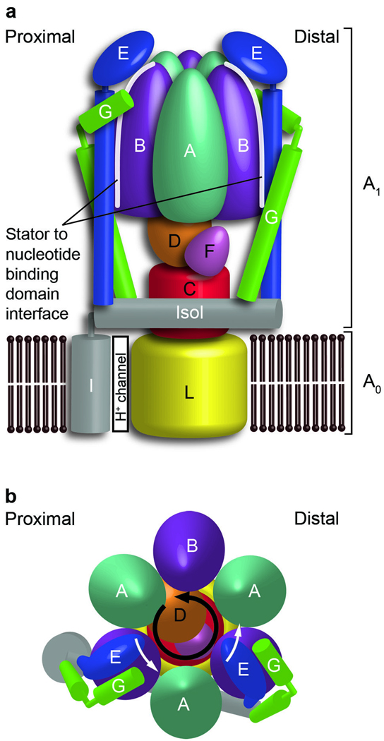
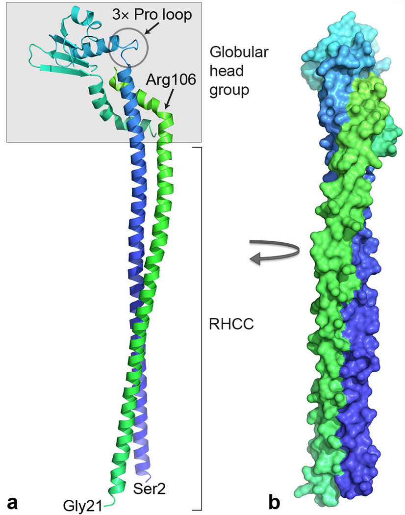
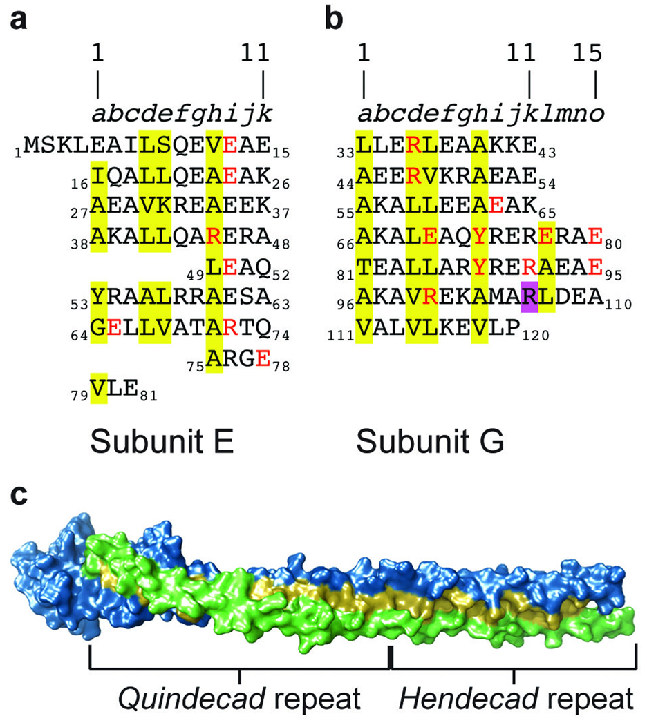
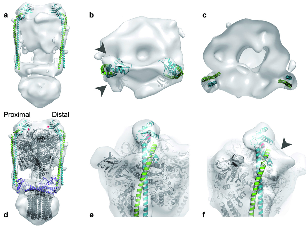
Proton-translocating ATPases are ubiquitous protein complexes that couple ATP catalysis with proton translocation via a rotary catalytic mechanism. The peripheral stalks are essential components that counteract torque generated from proton translocation during ATP synthesis or from ATP hydrolysis during proton pumping. Despite their essential role, the peripheral stalks are the least conserved component of the complexes, differing substantially between subtypes in composition and stoichiometry. We have determined the crystal structure of the peripheral stalk of the A-type ATPase/synthase from Thermus thermophilus consisting of subunits E and G. The structure contains a heterodimeric right-handed coiled coil, a protein fold never observed before. We have fitted this structure into the 23 A resolution EM density of the intact A-ATPase complex, revealing the precise location of the peripheral stalk and new implications for the function and assembly of proton-translocating ATPases.
Figures

Similar articles
-
The dynamic stator stalk of rotary ATPases.Nat Commun. 2012 Feb 21;3:687. doi: 10.1038/ncomms1693. Nat Commun. 2012. PMID: 22353718 Free PMC article.
-
Inter-subunit interaction and quaternary rearrangement defined by the central stalk of prokaryotic V1-ATPase.EMBO Rep. 2009 Nov;10(11):1228-34. doi: 10.1038/embor.2009.202. Epub 2009 Sep 25. EMBO Rep. 2009. PMID: 19779483 Free PMC article.
-
Structure of a central stalk subunit F of prokaryotic V-type ATPase/synthase from Thermus thermophilus.EMBO J. 2005 Nov 16;24(22):3974-83. doi: 10.1038/sj.emboj.7600859. Epub 2005 Nov 10. EMBO J. 2005. PMID: 16281059 Free PMC article.
-
Rotation, structure, and classification of prokaryotic V-ATPase.J Bioenerg Biomembr. 2005 Dec;37(6):405-10. doi: 10.1007/s10863-005-9480-1. J Bioenerg Biomembr. 2005. PMID: 16691473 Review.
-
Cryo-EM studies of the structure and dynamics of vacuolar-type ATPases.Sci Adv. 2016 Jul 22;2(7):e1600725. doi: 10.1126/sciadv.1600725. eCollection 2016 Jul. Sci Adv. 2016. PMID: 27532044 Free PMC article. Review.
Cited by
-
Reconstitution of vacuolar-type rotary H+-ATPase/synthase from Thermus thermophilus.J Biol Chem. 2012 Jul 13;287(29):24597-603. doi: 10.1074/jbc.M112.367813. Epub 2012 May 11. J Biol Chem. 2012. PMID: 22582389 Free PMC article.
-
Manipulations in the peripheral stalk of the Saccharomyces cerevisiae F1F0-ATP synthase.J Biol Chem. 2011 Mar 25;286(12):10155-62. doi: 10.1074/jbc.M110.213447. Epub 2011 Jan 21. J Biol Chem. 2011. PMID: 21257750 Free PMC article.
-
NMR solution structure of subunit E (fragment E(1-69)) of the Saccharomyces cerevisiae V (1)V (O) ATPase.J Bioenerg Biomembr. 2011 Apr;43(2):187-93. doi: 10.1007/s10863-011-9342-y. Epub 2011 Mar 12. J Bioenerg Biomembr. 2011. PMID: 21399923
-
A blue native-PAGE analysis of membrane protein complexes in Clostridium thermocellum.BMC Microbiol. 2011 Jan 26;11(1):22. doi: 10.1186/1471-2180-11-22. BMC Microbiol. 2011. PMID: 21269440 Free PMC article.
-
Biochemical and biophysical properties of interactions between subunits of the peripheral stalk region of human V-ATPase.PLoS One. 2013;8(2):e55704. doi: 10.1371/journal.pone.0055704. Epub 2013 Feb 11. PLoS One. 2013. PMID: 23409023 Free PMC article.
References
-
- Forgac M. Vacuolar ATPases: rotary proton pumps in physiology and pathophysiology. Nat. Rev. Mol. Cell Biol. 2007;8:917–929. - PubMed
-
- Yoshida M, Muneyuki E, Hisabori T. ATP synthase-a marvellous rotary engine of the cell. Nat. Rev. Mol. Cell Biol. 2001;2:669–677. - PubMed
-
- Cross RL, Muller V. The evolution of A-, F-, and V-type ATP synthases and ATPases: reversals in function and changes in the H+ATP coupling ratio. FEBS Lett. 2004;576:1–4. - PubMed
Publication types
MeSH terms
Substances
Associated data
- Actions
Grants and funding
LinkOut - more resources
Full Text Sources
Other Literature Sources
Molecular Biology Databases
