

Fiber delivered probe for efficient CARS imaging of tissues
- PMID: 20174068
- PMCID: PMC3014314
- DOI: 10.1364/OE.18.002380
Fiber delivered probe for efficient CARS imaging of tissues
Abstract
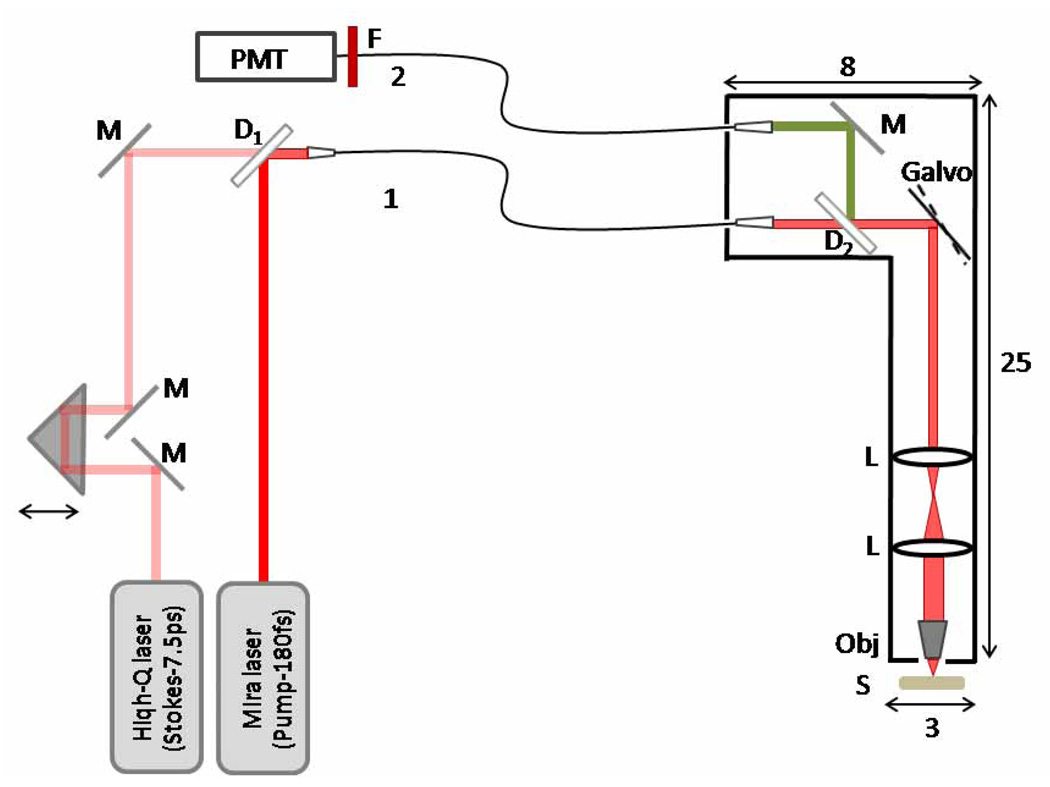
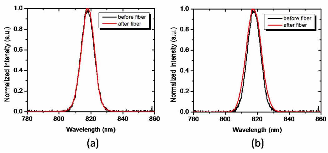
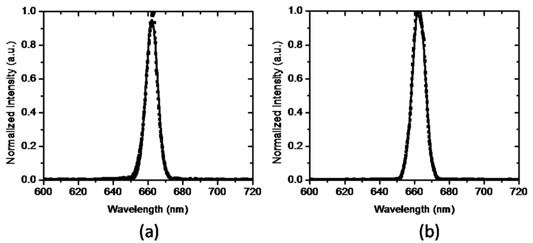
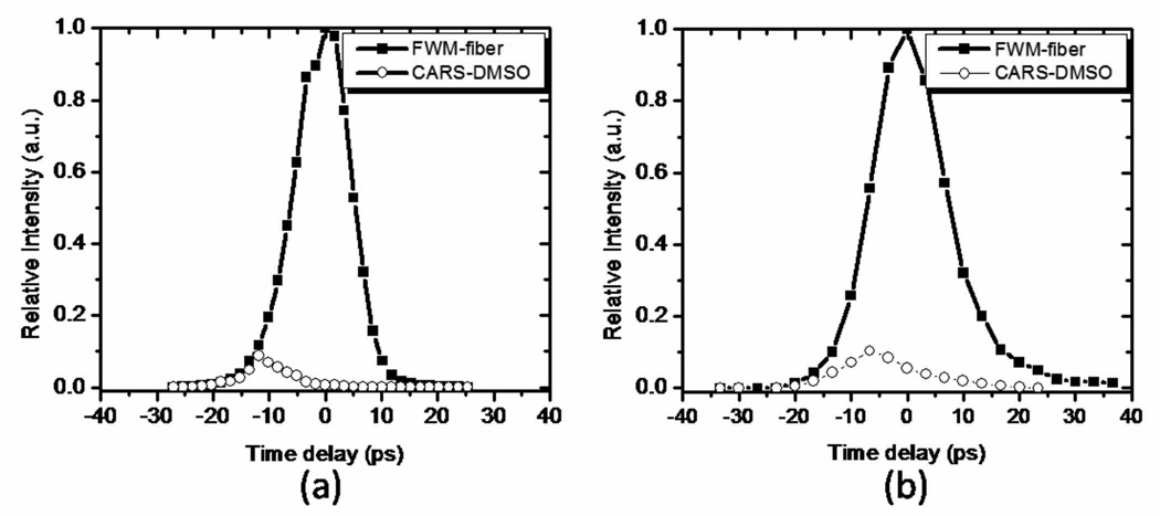
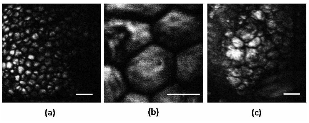
We demonstrate a fiber-based probe for maximum collection of the coherent anti-Stokes Raman scattering (CARS) signal in biological tissues. We discuss the design challenges including capturing the backscattered forward generated CARS signal in the sample and the effects of fiber nonlinearities on the propagating pulses. Three different single mode fibers (fused silica fiber, photonic crystal fiber and double-clad photonic crystal fiber) were tested for the probe design. We investigated self-phase modulation, stimulated Raman scattering (SRS) and four-wave-mixing (FWM) generation in the fiber: nonlinear processes expected to occur in a two-beam excitation based probe. While SPM and SRS induced spectral broadening was negligible, a strong non phase-matched FWM contribution was found to be present in all the tested fibers for excitation conditions relevant to CARS microscopy of tissues. To spectrally suppress this strong contribution, the pro design incorporates separate fibers for excitation light delivery and for signal detection, in combination with dichroic optics. CARS images of the samples were recorded by collecting the back-scattered forward generated CARS signal in the sample through a multi-mode fiber. Different biological tissues were imaged ex vivo in order to assess the performance of our fiber-delivered probe for CARS imaging, a tool which we consider an important advance towards label-free, in vivo probing of superficial tissues.
Figures
Similar articles
-
Coherent anti-Stokes Raman scattering microscopy imaging with suppression of four-wave mixing in optical fibers.Opt Express. 2011 Apr 25;19(9):7960-70. doi: 10.1364/OE.19.007960. Opt Express. 2011. PMID: 21643045
-
Use of multimode optical fibers for fiber-based coherent anti-Stokes Raman scattering microendoscopy imaging.Opt Lett. 2011 Aug 1;36(15):2967-9. doi: 10.1364/OL.36.002967. Opt Lett. 2011. PMID: 21808374
-
Investigation of a four-wave mixing signal generated in fiber-delivered CARS microscopy.Appl Opt. 2010 Jul 10;49(20):3916-21. doi: 10.1364/AO.49.003916. Appl Opt. 2010. PMID: 20648166
-
Label-free imaging of lipid dynamics using Coherent Anti-stokes Raman Scattering (CARS) and Stimulated Raman Scattering (SRS) microscopy.Curr Opin Genet Dev. 2011 Oct;21(5):585-90. doi: 10.1016/j.gde.2011.09.003. Epub 2011 Sep 22. Curr Opin Genet Dev. 2011. PMID: 21945002 Free PMC article. Review.
-
Coherent anti-stokes Raman scattering microscopy: a biological review.Cytometry A. 2006 Aug 1;69(8):779-91. doi: 10.1002/cyto.a.20299. Cytometry A. 2006. PMID: 16752420 Review.
Cited by
-
Mode-filtered large-core fiber for short-pulse delivery with reduced nonlinear effects.Opt Lett. 2011 Sep 1;36(17):3362-4. doi: 10.1364/OL.36.003362. Opt Lett. 2011. PMID: 21886211 Free PMC article.
-
Coherent anti-Stokes Raman scattering rigid endoscope toward robot-assisted surgery.Biomed Opt Express. 2018 Jan 2;9(2):387-396. doi: 10.1364/BOE.9.000387. eCollection 2018 Feb 1. Biomed Opt Express. 2018. PMID: 29552380 Free PMC article.
-
Chemical contrast for imaging living systems: molecular vibrations drive CARS microscopy.Nat Chem Biol. 2011 Mar;7(3):137-45. doi: 10.1038/nchembio.525. Nat Chem Biol. 2011. PMID: 21321552 Free PMC article. Review.
-
High-resolution multimodal flexible coherent Raman endoscope.Light Sci Appl. 2018 May 30;7:10. doi: 10.1038/s41377-018-0003-3. eCollection 2018. Light Sci Appl. 2018. PMID: 30839624 Free PMC article.
-
Beam scanning for rapid coherent Raman hyperspectral imaging.Opt Lett. 2015 Dec 15;40(24):5826-9. doi: 10.1364/OL.40.005826. Opt Lett. 2015. PMID: 26670522 Free PMC article.
References
-
- Evans CL, Xie XS. Coherent anti-Stokes Raman scattering microscopy: chemical imaging for biology and medicine. Annu. Rev. Anal. Chem. 2008;1:883–909. - PubMed
-
- Henry F, Côté D, Randolph MA, Rust EAZ, Redmond RW, Kochevar IE, Lin CP, Winograd JM. Real-time in vivo assessment of the nerve microenvironment with coherent anti-Stokes Raman scattering microscopy. Plastic and Reconstructive Surgery. 2009;123:123S–130S. - PubMed
Publication types
MeSH terms
Grants and funding
LinkOut - more resources
Full Text Sources
Other Literature Sources
