. 2009 Feb 1;2(2):85-120.
doi: 10.1007/s12274-009-9009-8.

Carbon Nanotubes in Biology and Medicine: In vitro and in vivo Detection, Imaging and Drug Delivery

Affiliations

Carbon Nanotubes in Biology and Medicine: In vitro and in vivo Detection, Imaging and Drug Delivery

Zhuang Liu et al. Nano Res. .

Abstract

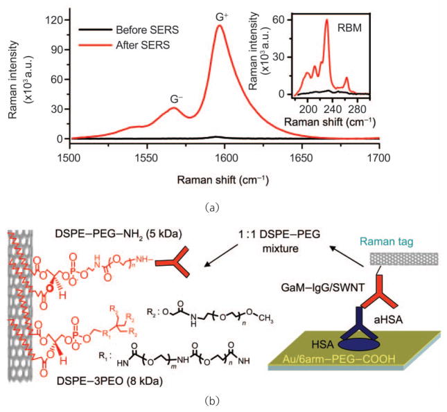
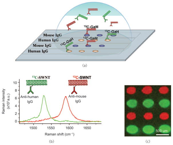
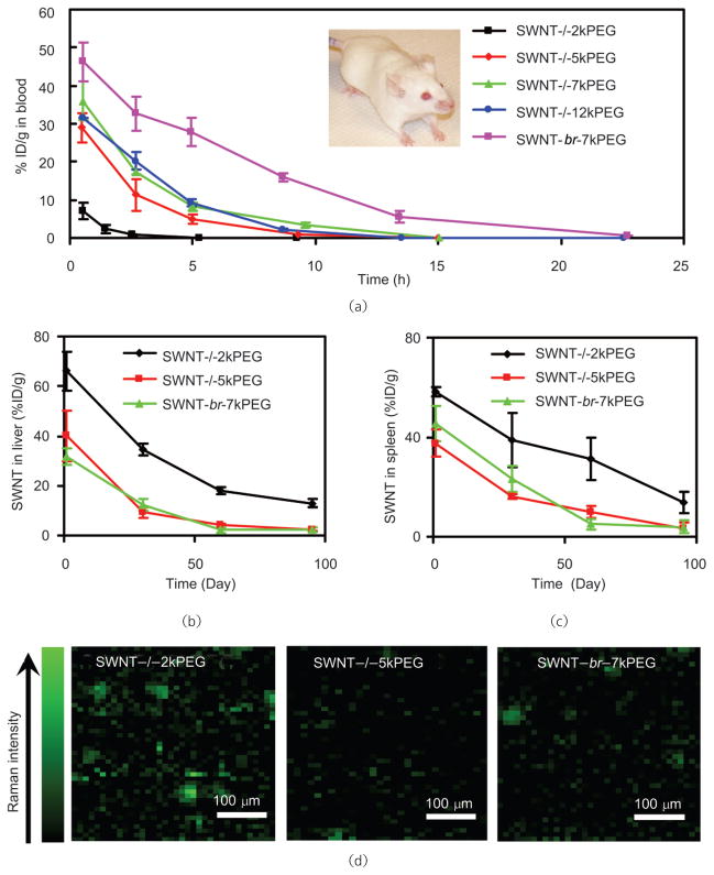
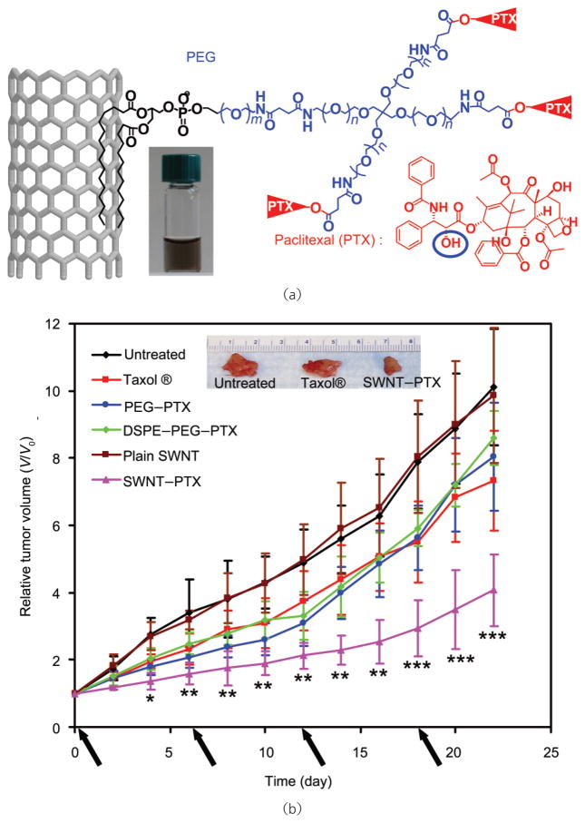
Carbon nanotubes exhibit many unique intrinsic physical and chemical properties and have been intensively explored for biological and biomedical applications in the past few years. In this comprehensive review, we summarize the main results from our and other groups in this field and clarify that surface functionalization is critical to the behavior of carbon nanotubes in biological systems. Ultrasensitive detection of biological species with carbon nanotubes can be realized after surface passivation to inhibit the non-specific binding of biomolecules on the hydrophobic nanotube surface. Electrical nanosensors based on nanotubes provide a label-free approach to biological detection. Surface-enhanced Raman spectroscopy of carbon nanotubes opens up a method of protein microarray with detection sensitivity down to 1 fmol/L. In vitro and in vivo toxicity studies reveal that highly water soluble and serum stable nanotubes are biocompatible, nontoxic, and potentially useful for biomedical applications. In vivo biodistributions vary with the functionalization and possibly also size of nanotubes, with a tendency to accumulate in the reticuloendothelial system (RES), including the liver and spleen, after intravenous administration. If well functionalized, nanotubes may be excreted mainly through the biliary pathway in feces. Carbon nanotube-based drug delivery has shown promise in various In vitro and in vivo experiments including delivery of small interfering RNA (siRNA), paclitaxel and doxorubicin. Moreover, single-walled carbon nanotubes with various interesting intrinsic optical properties have been used as novel photoluminescence, Raman, and photoacoustic contrast agents for imaging of cells and animals. Further multidisciplinary explorations in this field may bring new opportunities in the realm of biomedicine.


Figures

Figure 1
Figure 1
Optical properties of SWNTs. (a) Scheme of the electronic structure of SWNTs. The sharp features of the DOS are attributed to van Hove singularities. E11, E22, and E33 optical transitions correspond to photon absorption in the NIR, visible (vis) and UV ranges, respectively. (b) UV–vis–NIR absorption spectrum of an aqueous solution of SWNTs. Peaks in the spectrum are due to SWNTs with various chiralities. (c) Raman spectrum of SWNTs. The peaks at 200–300 cm−1, ~1400 cm−1, and ~1590 cm−1 are the radial breathing modes (RBM), D-band mode, and G-band mode, respectively. (d) Photoluminescence excitation (E22) and emission (E11) spectrum of semiconducting HIPCO SWNTs. SWNTs with different chiralities emit at various wavelengths under different excitations. The top panel illustrates the structures of three SWNT chiralities
Figure 2
Figure 2
Schemes of covalent functionalization of carbon nanotubes: (a) CNTs are oxidized and then conjugated with hydrophilic polymers (e.g., PEG) or other functional moieties; (b) photoinduced [1, 2] addition of azide compounds with CNTs; (c) Bingel reaction on CNTs; (d) 1,3-dipolar cylcoaddition on CNTs. For biological applications, “R” in the figure is normally a hydrophilic domain which renders CNTs water soluble. Further conjugation of bioactive molecules can be applied based on such functionalizations
Figure 3
Figure 3
Schemes of noncovalent functionalization of carbon nanotubes. (a) Proteins are anchored on the SWNT surface via pyrene π–π stacked on a nanotube surface. Right: A transmission electron microscope (TEM) image of an SWNT conjugated with proteins. Copyright 2001 American Chemical Society [70]. (b) An SWNT coated by a single-stranded DNA via π–π stacking. Copyright 2005 the National Academy of Sciences [20]. (c) An SWNT functionalized with PEGylated phospholipids. Both linear PEG (l-PEG) or branched PEG (br-PEG) can be used in this method. Copyright 2005 the National Academy of Sciences [45]
Figure 4
Figure 4
Tapping mode AFM images of the non-specific adsorption characteristics of streptavidin (SA) onto (a) bare single-walled carbon nanotubes (SWNTs) on SiO2/Si. (b) Adsorption of SA onto SWNTs and SiO2/Si following application of Triton X-100. Copyright 2002 American Chemical Society [83]
Figure 5
Figure 5
Specific chemical conjugation of biomolecules to SWNTs coated in Tween-20 surfactant, which prevents non-specific interactions, allows both quartz crystal microbalance (QCM) and FET measurement of target analyte binding. SWNTs functionalized by biotinylated Tween-20 (a) demonstrate QCM shifts dependent upon analyte, streptavidin, concentration (b), and analyte mediated conductance changes (c) as measured across a mat of SWNTs. (d) SWNTs may also be selectively coupled to Staphylococcal protein A (SpA) for selective binding of murine IgG as evidenced by QCM (e) and FET (f) measurements. Copyright 2003 the National Academy of Sciences [14]
Figure 6
Figure 6
SWNT network device architecture for increased Schottky contact area affords biosensors of increased sensitivity: (a) Shadow mask deposition of Au/Cr contacts at a 23° angle onto a semiconducting SWNT network increases Schottky contact area; (b) FET devices fabricated from SWNT networks with high Schottky contact area demonstrate enhanced sensitivities and limits of detection in specific biomolecule detection by four orders of magnitude. Copyright 2006 American Chemical Society [91]
Figure 7
Figure 7
SWNT-antibody conjugates may be utilized in conjunction with SERS as bright Raman tags for sandwich assay protein detection: (a) preparation of a spatially uniform SERS substrate for SWNT Raman tag detection of biomolecules significantly increases Raman scattering intensity, thus improving signal-to-noise and reducing assay time; (b) SWNT-antibody conjugates were formulated by first suspending SWNTs in aqueous media via PEGylated surfactants, which provide functionality and prevent non-specific interactions to the hydrophobic SWNT surface, and subsequently coupled to antibodies via bifunctional cross-linkers. Such antibody-Raman tags have been used in direct and indirect immunoassays. Copyright 2008 Nature Publishing Group [32]
Figure 8
Figure 8
SWNT-Raman tags have been applied to immunoassays demonstrating both high selectivity and sensitivity: (a) SWNT-anti-mouse IgG conjugates were applied to direct detection of mouse IgGs, demonstrating excellent signal-to-noise and minimal cross-reactivity; (b) SWNT-anti-mouse IgG conjugates were used as Raman tags for the indirect detection of mouse anti-human serum albumin (aHSA) IgG captured onto an SERS active substrate by HSA. A limit of detection of 1 fmol/L analyte was reproducibly observed (two separate trials are shown). The data are well fitted by a logistic regression (solid red curve, fit to data shown in green) allowing accurate quantitation of analyte over eight orders of magnitude. Copyright 2008 Nature Publishing Group [32]
Figure 9
Figure 9
SWNT tags made from pure 12C and 13C feedstocks may be used for multicolor Raman-based protein detection: (a) Schematic diagram of a protein microarray employing multi-color SWNT tags for high throughput, multiplexed detection; (b) 12C and 13C SWNTs demonstrate easily resolvable G-band Raman scattering spectra; (c) direct Raman detection of mouse IgG (red) and human IgG (green) by 12C and 13C SWNT tags, respectively. Copyright 2008 Nature Publishing Group [32]
Figure 10
Figure 10
Supramolecular chemistry of functionalized SWNTs for efficient drug loading and delivery. (a) Schematic of doxorubicin (DOX) π-stacking onto a nanotube pre-functionalized by PL-PEG. Targeting ligands such as RGD peptide can be conjugated on the PEG termini for targeted drug delivery. Inset: AFM images of SWNT before (top) and after (bottom) DOX loading. The height of SWNTs increased after DOX loading. (b) UV–vis–NIR absorbance spectra of solutions of free doxorubicin (green), plain SWNTs (black), and DOX loaded SWNTs. (c) Release curves of DOX from SWNTs at different pH. DOX loaded on SWNTs is stable at basic and neutral pH with very slow release but exhibits faster release in acidic environments. Copyright 2007 American Chemical Society [18]
Figure 11
Figure 11
siRNA delivery by carbon nanotubes: (a) a scheme of SWNT-siRNA conjugation via disulfide linkage; (b) confocal images of untreated cells (left) and SWNT–siRNACXCR4 treated cells (right) after PE-anti CXCR4 staining. Scale bars: 40 μm; (c) CXCR4 expression levels on CEM cells three days after various treatments, including four types of liposomes (Lipo1–4) and luciferase (Luc) siRNA control; (d) G-mode Raman intensity maps of single CEM cells after incubation for 1 d in SWNTs functionalized by PL-PEG2000 (left) and PL-PEG5400 (right) chains respectively. Inset: optical microscope images of CEM cells. Copyright 2007 Wiley-VCH [51]
Figure 12
Figure 12
In vivo pharmacokinetics and long-term biodistribution of SWNTs with different PEG coatings (Fig. 2(c)) measured by Raman spectroscopy. (a) Blood circulation curves of different PEGylated SWNTs after intravenous injection to mice. Long and branched PEG coating on SWNTs prolongs blood circulation half-lives of nanotubes. SWNT levels in liver (b) and spleen (c) over time. SWNTs are slowly excreted from RES organs in a few months, with faster clearance rate for those with good PEG coating. Note that SWNTs have much lower uptakes in other organs (less then 2% ID/g). (d) Raman images of mouse liver slices three months after SWNT injection. Much lower SWNT residues are observed in the liver of mice injected with nanotubes with more hydrophilic surface coating. Copyright 2008 the National Academy of Sciences [45]
Figure 13
Figure 13
In vivo tumor targeting with SWNTs. (a) Scheme of PEGylated SWNTs with RGD conjugation and radiolabeling. (b) Micro-PET images of mice. Arrows point to the tumors. High tumor uptake (~13 %ID/g) of SWNT–PEG5400–RGD is observed in the U87MG tumor (2nd column), in contrast to the low tumor uptake (1st column) of SWNT–PEG2000–RGD. The 3rd column is a control experiment showing blocking of SWNT–PEG5400–RGD tumor uptake by co-injection of free c(RGDyK). The 4th column is a control experiment showing low uptake of SWNT–PEG5400–RGD in integrin αvβ3-negative HT-29 tumor. Copyright 2007 Nature Publishing Group [44]
Figure 14
Figure 14
In vivo drug delivery with carbon nanotubes for cancer treatment. (a) Schematic illustration of paclitaxel conjugation to SWNT functionalized by phospholipids with branched-PEG chains. The PTX molecules are reacted with succinic anhydride (at the circled OH site) to form cleavable ester bonds and linked to the termini of branched PEG, via amide bonds. This allows for release of PTX from nanotubes by ester cleavage in vivo. (b) Tumor growth curves of 4T1 tumor-bearing mice receiving the different treatments indicated. The same PTX dose (5 mg/kg) was injected (on days 0, 6, 12, and 18, marked by arrows) for Taxol®, PEG–PTX, DSEP–PEG–PTX and SWNT–PTX. p Values (Taxol® vs SWNT–PTX): * p<0.05, ** p<0.01, *** p<0.001. Inset: A photo of representative tumors taken out of an untreated mouse, a Taxol® treated mouse and an SWNT–PTX treated mouse at the end of the treatments. Copyright 2008 American Association for Cancer Research [37]
Figure 15
Figure 15
SWNT as NIR fluorescent labels. (a) Schematic of targeting cells with SWNT-Herceptin conjugates to BT-474 (HER2/neu positive) and MCF-7 (HER2/neu negative) cells. (b) NIR photoluminescence image of BT-474 cells treated with the SWNT-herceptin conjugate. The high level of fluorescence signal indicates surface binding of the SWNT-Herceptin conjugate. (c) NIR photoluminescence image of MCF-7 cells. Very little NIR signal is seen owing to the low non-specific binding of the SWNT-Herceptin conjugate and low cellular autofluorescence. NIR photoluminescence images were taken using a homebuilt scanning confocal microscope with InGaAs detector. Photoluminescence was collected from 900–1600 nm. Copyright 2008 American Chemical Society [24]
Figure 16
Figure 16
Multi-color Raman imaging with isotopically modified SWNTs. (a) Schematic illustration of SWNTs with three different isotope compositions (13C-SWNT, 12C12/13C-SWNT, 12C-SWNT) conjugated with different targeting ligands. (b) Solution phase Raman spectra of the three SWNT conjugates under 785 nm laser excitation. Different G-band peak positions were observed. (c) A deconvoluted confocal Raman spectroscopy image of a mixture of three cell lines with different receptor expressions incubated with the three-color SWNT mixture. Scale bar = 100 μm. Copyright 2008 American Chemical Society [127]
Figure 17
Figure 17
In vivo tumor imaging with SWNTs. In vivo photoacoustic (strong light absorption of SWNTs generates an acoustic signal) and Raman images of U87MG tumors in live mice injected with plain SWNT or RGD-conjugated SWNT. Strong tumor contrast induced by SWNT–RGD was observed in both imaging techniques. Copyright 2008 American Chemical Society [154], 2008 Nature Publishing Group [22], and 2008 the National Academy of Sciences [153]

References

    1. Whitesides GM. The “right” size in nanobiotechnology. Nat Biotech. 2003;21:1161–1165. - PubMed
    1. Lowe CR. Nanobiotechnology: The fabrication and applications of chemical and biological nanostructures. Curr Opin Chem Biol. 2000;10:428–434. - PubMed
    1. Wang L, Zhao W, Tan W. Bioconjugated silica nanoparticles: Development and applications. Nano Res. 2008;1:99–115.
    1. Iijima S. Helical microtubules of graphitic carbon. Nature. 1991;354:56–58.
    1. Dai H. Carbon nanotubes: Synthesis, integration, and properties. Acc Chem Res. 2002;35:1035–1044. - PubMed

LinkOut - more resources