. 2010 Feb 19;6(2):e1000852.
doi: 10.1371/journal.pgen.1000852.

Proteasome nuclear activity affects chromosome stability by controlling the turnover of Mms22, a protein important for DNA repair

Affiliations

Proteasome nuclear activity affects chromosome stability by controlling the turnover of Mms22, a protein important for DNA repair

Shay Ben-Aroya et al. PLoS Genet. .

Abstract

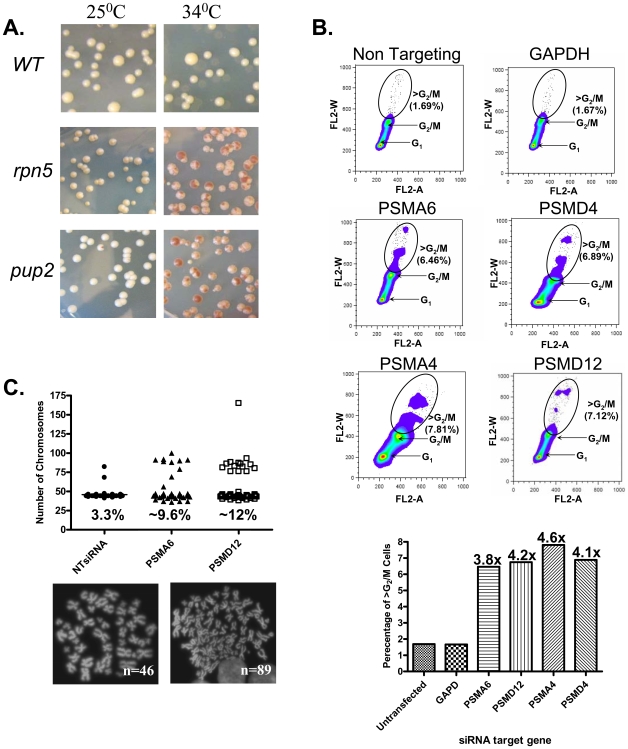
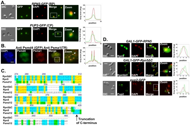
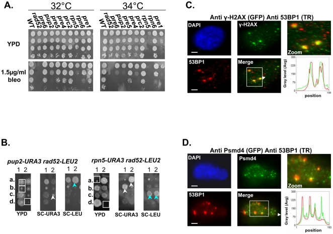
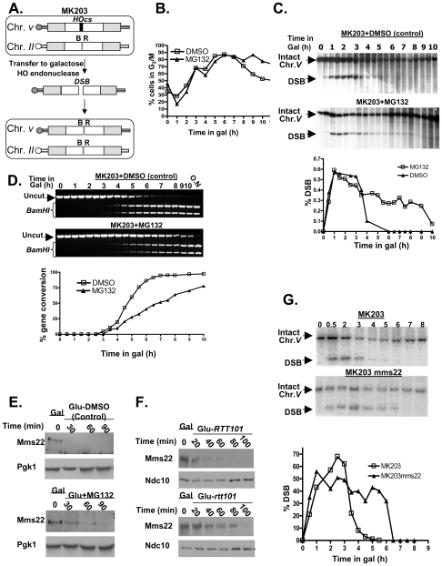
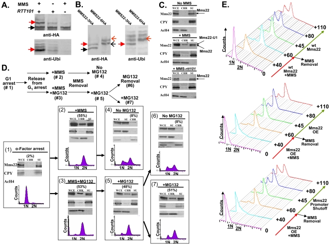
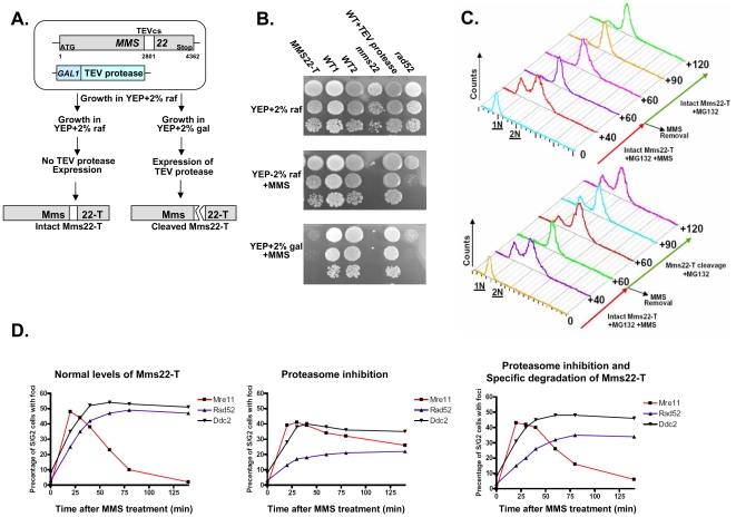
To expand the known spectrum of genes that maintain genome stability, we screened a recently released collection of temperature sensitive (Ts) yeast mutants for a chromosome instability (CIN) phenotype. Proteasome subunit genes represented a major functional group, and subsequent analysis demonstrated an evolutionarily conserved role in CIN. Analysis of individual proteasome core and lid subunit mutations showed that the CIN phenotype at semi-permissive temperature is associated with failure of subunit localization to the nucleus. The resultant proteasome dysfunction affects chromosome stability by impairing the kinetics of double strand break (DSB) repair. We show that the DNA repair protein Mms22 is required for DSB repair, and recruited to chromatin in a ubiquitin-dependent manner as a result of DNA damage. Moreover, subsequent proteasome-mediated degradation of Mms22 is necessary and sufficient for cell cycle progression through the G(2)/M arrest induced by DNA damage. Our results demonstrate for the first time that a double strand break repair protein is a proteasome target, and thus link nuclear proteasomal activity and DSB repair.


Conflict of interest statement

The authors have declared that no competing interests exist.

Figures

Figure 1
Figure 1. The CIN Phenotype of proteasome subunits is conserved from yeast to human cells.
(A) Chromosome transmission fidelity (ctf) phenotype of yeast mutants defective for the proteasome subunits pup2 and rpn5ΔC at a semi-permissive temperature (34°C) is scored by the appearance of sectored colonies, and compared to the isogenic wt strain. (B) DNA content dot plots of asynchronous HCT116 cells following siRNA knockdown in control, and test cases generated from cell populations harvested 5-days after transfection. HCT116 cell line, a mismatch repair-deficient cell line, was used, as it is a chromosomally stable, near diploid colorectal cell line that does not inherently exhibit CIN. Cells were labeled with propidium iodide and subjected to flow cytometry. Circles delineate the population of cells having >G2/M DNA contents. The graph summarizes the relative increase in this cell population as compared to the non-targeting and GAPDH controls. (C) Scatter plot depicting the total chromosome number distribution after targeted knockdown of PSMA6, PSMD12, or a non targeting (NT) RNAi control. Percentage of mitotic spreads with greater than 46 chromosomes is indicated at the base of each column; (below) Representative images of DAPI-stained mitotic spreads from untransfected cells (N = 46 chromosomes) and aneuploid cells after treatment with PSMA6 siRNA (N = 89 chromosomes).
Figure 2
Figure 2. Proteasome subunits in yeast and mammalian cells localize to the nucleus; the Ts allele of rpn5 is truncated at the C-terminus; proteasome CIN mutants show nucleus mislocalization of proteasome subunits.
(A,B) Proteasome subunits in yeast and mammalian cells localize to the nucleus. (C) The Ts allele of rpn5 is truncated at the C-terminus. (D) Proteasome CIN mutants show nucleus mislocalization of proteasome subunits. The panels represent high resolution (x100) representative images of yeast or mammalian cells. A region identified by the white box is further magnified (zoom panel). The position of the white arrow within the zoom panel delineates the line scan that was used to quantitate the fluorescent signal intensities per pixel in the line scan graphs (right panel). Unless otherwise stated, all the images represent a 3-D projection of x100 Z-series images extending above and below the entire nucleus. Scale bars, 3 µm. (A) Logarithmic yeast cultures were permeabilized and DNA was DAPI stained to mark the nucleus. The panels depict the nuclear localization of the yeast Regulatory Particle (RP) Rpn5-GFP, and Core Particle (CP) Pup2-GFP. GFP and DAPI are represented by green and red curves in the line scan graphs, respectively. (B) Nuclear enrichment and foci colocalization of immunofluorescently labeled mammalian proteasomal subunits Psma1 (CP) and Psmd4 (RP). Cells were DAPI stained and visualized by GFP, Texas Red (TR) and DAPI. Red and green lines represent TR and GFP, respectively. Panels represent a 3-D projection of x100 Z-series images extending above and below the entire nucleus. (C) The rpn5-Ts allele was sequenced and its predicted translation product aligned to the wt yeast protein Rpn5, and its human homolog, Psmd12. The truncation point of Rpn5ΔC is indicated by a black arrow. (D) Localization analysis of N-terminal GFP fusion of Rpn5ΔC, and Rpn5 control (both expressed from a galactose-inducible promoter). The images depict the localization of GAL1-GFP-Rpn5 vs. GAL1-GFP-Rpn5ΔC. GFP-Rpn5 localizes to the nucleus (overlap between the DAPI and GFP channels). Lack of overlap in GFP-Rpn5ΔC indicates nuclear mislocalization. Similar results were obtained for pup2-Ts (Pup2-GFP) (compare to Figure 2A).
Figure 3
Figure 3. Mutated proteasome subunits affect the repair of DNA DSBs.
(A) Most of proteasomal Ts mutants are sensitive to bleomycin (bleo). Five-fold serial dilutions of the indicated proteasomal subunits mutants were spotted on YPD medium lacking or supplemented with 1.5 µ/ml of bleo. Cells were incubated at 32°C and 34°C to find the semi-permissive temperature of each Ts mutant. (B) rpn5ΔC and pup2 show synthetic growth defect with rad52. To examine whether there could be a link between the proteasome and the repair of DSBs, we created and sporulated heterozygous diploid strains containing Ts alleles of either rpn5ΔC and pup2 combined with rad52. Tetrad dissection showed that Ts alleles of rpn5ΔC and pup2 cause a synthetic growth defect when either one is combined with rad52. The synthetic growth defect of the double mutant spores (encircled by white squares on the YPD plate) is evident when compared to the single haploid mutants (pointed out by white or light blue arrows). (C,D) Protesomal subunits associate with DSB markers in mammalian cells. HeLa cells were treated for 2 hrs with 5 µ/ml of bleo prior to subjection to IIF microscopy. Primary antibodies were recognized with appropriate secondary antibodies conjugated with either Alexa-fluor 488 (GFP filter), or Cy-3 (TR filter). Scale bars, 3 µm. (C) IIF to demonstrate the colocalization pattern of 53BP1 and γ-H2AX in bleo-treated cells. The DSB markers 53BP1 and γ-H2AX show clear co-localization at large foci, likely to represent DSB sites. Red and green curves on the line scan graph represent 53BP1, and γ-H2AX respectively. (D) Representative images demonstrating an association of the RP subunit Psmd4 with DSB sites, represented by the large 53BP1 foci. Red and green curves on the line scan graph represent 53BP1 and Psmd4 respectively.
Figure 4
Figure 4. Proteasome mutants exhibit defective DSB repair kinetics; the turn over of Mms22 is regulated by the proteasome; Mms22 plays a role in DSB repair.
(A–D) Proteasome mutants exhibit defective DSB repair kinetics. (E,F) The turn over of Mms22 is regulated by the proteasome. (G) Mms22 plays a role in DSB repair. (A) Schematic representation of MK203 (for more details see Materials and Methods, DSB repair kinetic experiments). White rectangles represent the ura3 alleles on chromosomes II and V. A black bar within the ura3 alleles represents the HO cut site (HOcs); a grey bar depicts the inactive HOcs-inc flanked by the BamHI (B) and EcoRI (R) restriction sites. Transfer of the cells to galactose-containing medium results in a DSB that is repaired by homologous recombination. (B–D) DSB repair kinetics of MK203 cells in the presence of proteasome inhibitor. MK203 pdr5 cells were grown to mid-logarithmic phase in glycerol-containing medium (gly) (no HO-induction) containing 20 mM MG132, or DMSO control. Cells were then transferred to galactose-containing medium (gal; constitutive HO-induction and DSB formation at the URA3 locus) containing the same concentration of the drug. Samples were collected for analysis at timely intervals, and subjected to microscopic examination and Southern blot analysis. (B) Microscopic examination of dumbbell shaped cells indicates the percentage of G2/M in the control, or cells subjected to MG132, at each of the indicated time points. (C) Southern blot analysis and quantification graph (bottom) of the DSB repair kinetics in MK203 cells treated with MG132. (D) PCR analysis of the kinetics of the gene conversion product formation. PCR reaction followed by BamHI restriction digest detect the final step of the repair, which is the re-ligation of the broken ends and transfer of the two polymorphic restriction sites on either side of the HOcs from chromosome II to chromosome V . MG132 treated cells show a delay in gene conversion product formation, as apparent from the quantification graph (bottom). (E) Western Blot detects the levels of Mms22 following a GAL1 promoter shut-off chase experiment. The expression of GAL1-HA-Mms22 was induced by growing the cells in 2% galactose (Gal) for 3 hours (t-0). Cells were released into 2% glucose to shut-off the expression of Mms22. Glucose was supplemented with 20 mM MG132, or with DMSO (control), Pgk1 was used as a loading control. (F) RTT101 regulates the levels of Mms22. GAL1 shut-off chase experiment was performed as in (E), this time wt cells vs. rtt101 strains were released into 2% glucose. Ndc10 was used as a loading control. (G) Southern blot analysis and quantification graph (bottom) of the DSB repair kinetics in wt MK203 cells versus mms22.
Figure 5
Figure 5. Ubiquitination of Mms22 is induced by DNA damage in a RTT101 dependent manner; Mms22 is recruited to chromatin upon DNA damage in a RTT101 dependent manner; degradation of Mms22 from chromatin is associated with exit from the DNA damage induced G2/M arrest.
(A, B) Ubiquitination of Mms22 is induced by DNA damage in a RTT101 dependent manner. (C) Mms22 is recruited to chromatin upon DNA damage in a RTT101 dependent manner. (D,E) Degradation of Mms22 from chromatin is associated with exit from the DNA damage induced G2/M arrest. (A) 3HA-tagged Mms22 cells were grown in the presence of 20mM MG132, with or without 0.025% MMS, and subjected to IP. After electrophoresis on a low percentage gel (6%), the precipitated protein was blotted to a membrane which was successively immunobloted with an anti-HA, and an anti-Ubi antibody. Black arrow labels Mms22-3HA, red arrow labels the modified form of Mms22-3HA. (B) In vivo demonstration that the ubiquitinated proteins observed in (A). are a series of polyubiquitinated forms of Mms22. Cells carrying Mms22 tagged with either 3HA, or 6HA were grown in the presence of 20 mM MG132, and 0.025% MMS, and subjected to IP. After electrophoresis the precipitated proteins were blotted to membranes which were subjected to immunoblotting with anti-HA, and anti-Ubi antibodies. Black and open black arrows label Mms22-3HA and Mms22-6HA respectively. Red and open red arrows label the mono-ubiquitinated form of Mms22-3HA and Mms22-6HA respectively. (C) Cell extracts (WCE) were separated into supernatant (SU) and chromatin (CH) fractions. HA-tagged Mms22 was detected by immunoblotting in wt (middle), or rtt101 deleted cells (right), treated with 0.025% MMS, and compared to the untreated wt control (left). Anti Carboxy peptidase-Y (CPY), and Anti Acetylated Histon H4 (AcH4) served as a SU and CH fractions controls respectively. (D) (Top)-Experimental design. Chromatin fractionation assay was performed as in (B). The quantity of Mms22 on the chromatin bound fraction is represented as percentage of the WCE. Cells were synchronized to G1 (#1), and released from the arrest in the presence of 0.025% MMS (#2), or MMS+MG132 (#3). Next, MMS was removed, and samples were allowed to recover in the presence (#5), or absence (#6) of MG132. Sample #6 and #7 represent a division of sample #6 to a sample without, or with MG132 respectively. (E) Failure to degrade Mms22 impairs progression of repair, leading to prolonged cell cycle arrest. G1 arrested cells were released into YEP-Gal medium (inducing overexpression of GAL1-MMS22 cells) containing 0.025% MMS. MMS was washed from the G2/M arrested cells, and cells were allowed to recover in YEP-Gal or YEP-Glu (thus keeping either high or low expression levels of Mms22 in GAL1-MMS22 cells respectively). Top panel: wt expression levels of Mms22. Middle panel: Mms22 was over-expressed (OE) before, and after the removal of MMS. Bottom panel: Mms22 was OE before the removal of MMS, while its GAL1 promoter was shut off following MMS removal. Samples were collected at timely intervals and subjected to FACs analysis; numbers along the red and green arrows represent the time (minutes) since the release from the G1 arrest, or following MMS removal respectively.
Figure 6
Figure 6. Degradation of Mms22 is sufficient to allow exit from the DNA damage induced G2/M arrest.
(A,B) Schematic representation of the experimental design, and growth phenotypes. The TEV protease consensus cleavage site (cs) was introduced at position 2801 of Mms22 which is expressed from its endogenous promoter (MMS22-T). The cells also contain the TEV protease under the control of the inducible GAL promoter . YEP medium supplemented with 2% raffinose (raf) suppresses the expression of the TEV protease, and keeps Mms22-T functional, as indicated by its normal growth on YEP+raf+MMS media (compare to mms22 strain). Transfer of the cells to medium containing 2% galactose (gal) results in TEV protease induction, and the specific cleavage of Mms22-T. The inactivation of Mms22-T is indicated by its impaired growth on YEP+gal+MMS medium. (C) G1 arrested cells were released into YEP-raf medium (which blocks the expression of the TEV-protease), containing 0.025% MMS and 20 mM MG132. MMS was then washed from the G2/M arrested cells, and cells were allowed to recover in YEP-raf (top), or YEP-gal (bottom) (intact, or specific cleavage of Mms22-T respectively), both supplemented with MG132. Samples were collected at timely intervals and subjected to FACS analysis. Numbers along the red and green arrows represent the time (minuets) since the release from the G1 arrest, or following the removal of MMS respectively. (D) Temporal analysis of Mre11, Ddc2, and Rad52 focus formation following DNA damage and proteasome inhibition. Yeast strains containing Mms22-T, GAL inducible TEV-protease, and a YFP tagged version of either Mre11, Ddc2, or Rad52 were released into non-inducing YEP-raf medium containing 0.025% MMS and 20 mM MG132 (intact Mms22-T). Following the induction of DNA damage MMS was washed from the media, and cells were allowed to recover in YEP-raf (left: wt levels of Mms22), YEP-raf+MG132 (middle: accumulation of intact Mms22-T), or YEP-gal+MG132 (right: inducing the specific cleavage of Mms22-T by the TEV protease). Following the removal of MMS, samples were collected at timely intervals, fixed, and subjected to fluorescent microscopy. At each of the indicated time points at least 150 S/G2 cells (budded) were analyzed for the presence/absence of Mre11, Ddc2, or Rad52 foci.

Similar articles

Cited by

References

    1. Lengauer C, Kinzler KW, Vogelstein B. Genetic instabilities in human cancers. Nature. 1998;396:643–649. - PubMed
    1. Myung K, Kolodner RD. Suppression of genome instability by redundant S-phase checkpoint pathways in Saccharomyces cerevisiae. Proc Natl Acad Sci U S A. 2002;99:4500–4507. - PMC - PubMed
    1. Branzei D, Foiani M. Interplay of replication checkpoints and repair proteins at stalled replication forks. DNA Repair (Amst) 2007;6:994–1003. - PubMed
    1. Kolodner RD, Putnam CD, Myung K. Maintenance of genome stability in Saccharomyces cerevisiae. Science. 2002;297:552–557. - PubMed
    1. Symington LS. Role of RAD52 epistasis group genes in homologous recombination and double-strand break repair. Microbiol Mol Biol Rev. 2002;66:630–670, table of contents. - PMC - PubMed

Publication types

MeSH terms