. 2010 Feb 19;5(2):e9298.
doi: 10.1371/journal.pone.0009298.

Variable Na(v)1.5 protein expression from the wild-type allele correlates with the penetrance of cardiac conduction disease in the Scn5a(+/-) mouse model

Affiliations

Variable Na(v)1.5 protein expression from the wild-type allele correlates with the penetrance of cardiac conduction disease in the Scn5a(+/-) mouse model

Anne-Laure Leoni et al. PLoS One. .

Abstract

Background: Loss-of-function mutations in SCN5A, the gene encoding Na(v)1.5 Na+ channel, are associated with inherited cardiac conduction defects and Brugada syndrome, which both exhibit variable phenotypic penetrance of conduction defects. We investigated the mechanisms of this heterogeneity in a mouse model with heterozygous targeted disruption of Scn5a (Scn5a(+/-) mice) and compared our results to those obtained in patients with loss-of-function mutations in SCN5A.

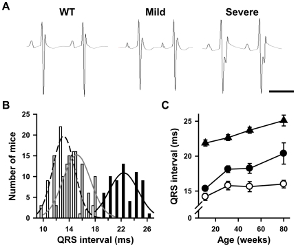
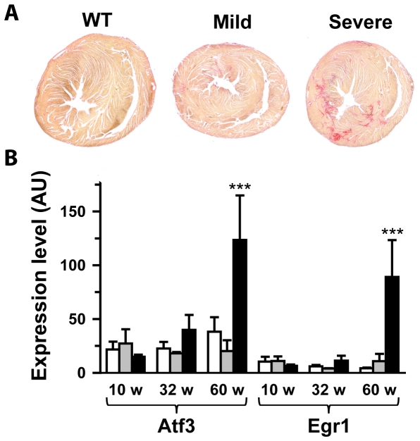
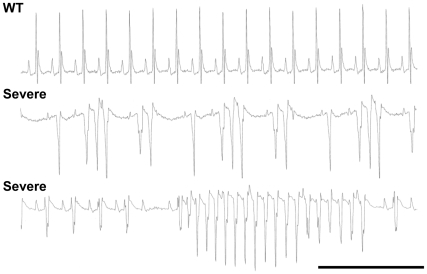
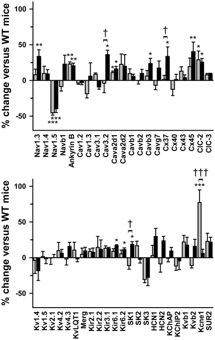
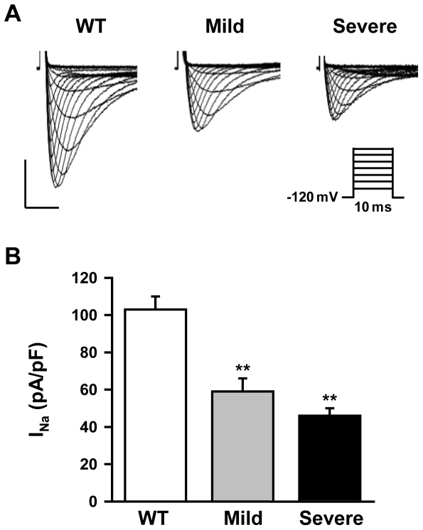
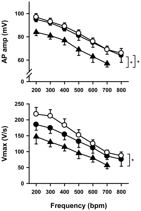
Methodology/principal findings: Based on ECG, 10-week-old Scn5a(+/-) mice were divided into 2 subgroups, one displaying severe ventricular conduction defects (QRS interval>18 ms) and one a mild phenotype (QRS< or = 18 ms; QRS in wild-type littermates: 10-18 ms). Phenotypic difference persisted with aging. At 10 weeks, the Na+ channel blocker ajmaline prolonged QRS interval similarly in both groups of Scn5a(+/-) mice. In contrast, in old mice (>53 weeks), ajmaline effect was larger in the severely affected subgroup. These data matched the clinical observations on patients with SCN5A loss-of-function mutations with either severe or mild conduction defects. Ventricular tachycardia developed in 5/10 old severely affected Scn5a(+/-) mice but not in mildly affected ones. Correspondingly, symptomatic SCN5A-mutated Brugada patients had more severe conduction defects than asymptomatic patients. Old severely affected Scn5a(+/-) mice but not mildly affected ones showed extensive cardiac fibrosis. Mildly affected Scn5a(+/-) mice had similar Na(v)1.5 mRNA but higher Na(v)1.5 protein expression, and moderately larger I(Na) current than severely affected Scn5a(+/-) mice. As a consequence, action potential upstroke velocity was more decreased in severely affected Scn5a(+/-) mice than in mildly affected ones.

Conclusions: Scn5a(+/-) mice show similar phenotypic heterogeneity as SCN5A-mutated patients. In Scn5a(+/-) mice, phenotype severity correlates with wild-type Na(v)1.5 protein expression.


Conflict of interest statement

Competing Interests: The authors have declared that no competing interests exist.

Figures

Figure 1
Figure 1. Variable degrees of ventricular conduction defects in Scn5a +/− mice.
A. Representative lead I ECGs from 10 week-old wild-type (WT) mice and Scn5a +/− mice with mild and severe conduction defects. Scale bar, 100 ms. B. Distribution of QRS interval duration with corresponding Gaussian fits in 10 week-old WT mice (white bars) and Scn5a +/− mice with mild (grey bars) and severe (black bars) phenotype. C. Effects of age (X-axis) on QRS interval duration (Y-axis) in WT mice (open symbols) and Scn5a +/− mice with mild (filled circles) and severe (filled triangles) phenotype. See Table 1 for statistics.
Figure 2
Figure 2. Variable effects of Na+ channel blockade in Scn5a +/− mice and SCN5A-mutated patients.
Ajmaline-induced increase in QRS interval (Y-axes) in 10 week- and >53 week-old WT (n = 11 and n = 10; open bars), mildly (n = 10 and n = 10; grey bars) and severely (n = 11 and n = 10; black bars) affected Scn5a +/− mice (left panel), and in young (6–31 years; mean = 22 years) and older (41–61 years; mean = 50 years) non-mutated (n = 18 and n = 17, open bars) and SCN5A-mutated patients with either mild (n = 12 and n = 11; grey filled bars) or severe (n = 7 and n = 12; black filled bars) QRS interval prolongation (right panel). *, ***, p<0.05 and p<0.001 respectively versus WT mice or non-mutated patients. †, p<0.05 versus Scn5a +/− mice or SCN5A-mutated patients with a mild phenotype. The difference between old mildly affected mice and old WT mice did not reach significance (p = 0.07). Same comment for the older patients.
Figure 3
Figure 3. Variable levels of fibrosis in Scn5a +/− mice.
A. Sirius red staining of ventricle from 85 week-old WT, mildly and severely affected Scn5a +/− mice. Fibrosis appears in red. A score of 0 was attributed to the WT mouse shown, 1 to the mild Scn5a +/− mouse and respectively 2 and 3 to left and right severe Scn5a +/− mice. B. Atf3 and Egr1 mRNA ventricular levels (in arbitrary units) in WT (open bars), mildly (grey bars) and severely (black bars) affected Scn5a +/− mice as a function of age. ***, p<0.001 versus WT and mild.
Figure 4
Figure 4. Cardiac arrhythmias in Scn5a +/− mice with severe phenotype.
Representative episodes of spontaneous ventricular arrhythmias recorded in two different Scn5a +/− mice with a severe phenotype under anesthesia. An ECG recorded in an age-matched WT mouse is given for comparison. Scale bar, 500 ms.
Figure 5
Figure 5. Moderate ionic remodeling in Scn5a +/− mice.
Percentage of variation in ventricular expression (Y-axes) of 46 genes encoding ion channel subunits (ch) and connexins (Cx) in Scn5a +/− mice with mild (grey bars; n = 5) and severe (black bars; n = 7) phenotype versus WT mice (n = 10). Sub, subunits. *, **, ***, p<0.05, p<0.01 and p<0.001 respectively versus WT; †, †††, p<0.05 and p<0.001 respectively versus mild phenotype.
Figure 6
Figure 6. Decreased Nav1.5 expression in Scn5a +/− mice.
A. Representative Western blots showing the expression levels of Nav1.5 in WT mice and Scn5a +/− mice (mean age = 18±1 weeks) with a mild or a severe phenotype. Protein loading was controlled by anti-actin immunoblotting. B. Quantification of Nav1.5 expression in WT mice (open bars) and Scn5a +/− mice with mild (grey bars) and severe (black bars) phenotype was performed on Western blots from 10 mice in each group by normalizing the intensities of the Nav1.5 bands to the actin bands. ***, p<0.001 versus WT; †, p<0.05 versus mild Scn5a +/− mice. C. Confocal images of Nav1.5 in ventricular cardiomyocytes isolated from WT mice (left panel) and Scn5a +/− mice with mild (middle panel) and severe (right panel) phenotype. Scale bar, 20 µm. In Scn5a +/− images presented, a WT cardiomyocyte is also included in the image frame (yellow arrows) to illustrate the clear difference in WT versus Scn5a +/− intercalated disc staining intensities.
Figure 7
Figure 7. Variable INa densities in Scn5a +/− single ventricular cardiomyocytes.
A. Representative INa traces (protocol in inset) obtained from ventricular myocytes in a 12 week-old WT mouse and Scn5a +/− mice with mild and severe phenotype. Horizontal bar, 2 ms; vertical bar, 40 pA/pF. B. INa density in myocytes from WT and Scn5a +/− mice with mild and severe phenotype (4 mice in each group, 4 to 6 cells for each mouse). **, p<0.01 versus WT mice.
Figure 8
Figure 8. Action potential amplitude (AP amp; top) and maximum upstroke velocity (Vmax; bottom) as a function of pacing frequency.
White circles, wild type mice (n = 7); black circles, Scn5a +/− mice with a mild phenotype (n = 6); black triangles, Scn5a +/− mice with a severe phenotype (n = 5). At 800 bpm, 4 preparations from Scn5a +/− mice with a severe phenotype were periodically (for 1 of them) or continuously (for the 3 others) in 2∶1 conduction block. *, p<0.05.

References

    1. Schott J-J, Alshinawi C, Kyndt F, Probst V, Hoorntje TM, et al. Cardiac conduction defects associate with mutations in SCN5A. Nat Genet. 1999;23:20–21. - PubMed
    1. Probst V, Kyndt F, Potet F, Trochu J-N, Mialet G, et al. Haploinsufficiency in combination with aging causes SCN5A-linked hereditary Lenegre disease. J Am Coll Cardiol. 2003;41:643–652. - PubMed
    1. Chen Q, Kirsch GE, Zhang D, Brugada R, Brugada J, et al. Genetic basis and molecular mechanism for idiopathic ventricular fibrillation. Nature. 1998;392:293–296. - PubMed
    1. Tan HL, Bezzina CR, Smits JP, Verkerk AO, Wilde AA. Genetic control of sodium channel function. Cardiovasc Res. 2003;57:961–973. - PubMed
    1. Clancy CE, Kass RS. Defective cardiac ion channels: from mutations to clinical syndromes. J Clin Invest. 2002;110:1075–1077. - PMC - PubMed

Publication types

MeSH terms