. 2010 Feb 18;5(2):e9299.
doi: 10.1371/journal.pone.0009299.

Skeletal muscle phenotypically converts and selectively inhibits metastatic cells in mice

Affiliations

Skeletal muscle phenotypically converts and selectively inhibits metastatic cells in mice

Ara Parlakian et al. PLoS One. .

Abstract

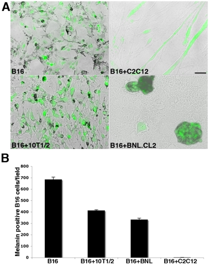
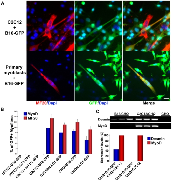
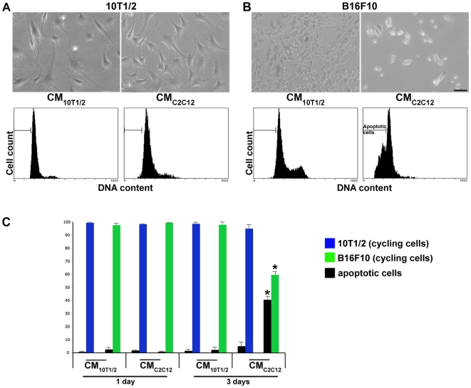
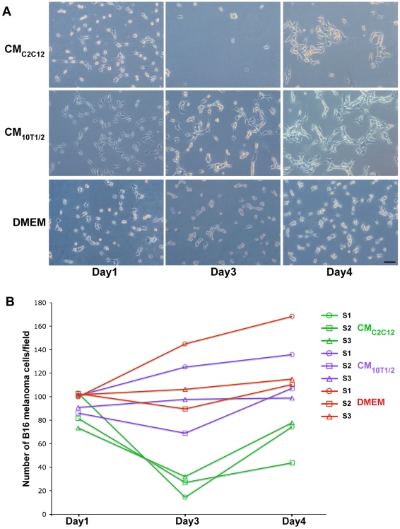
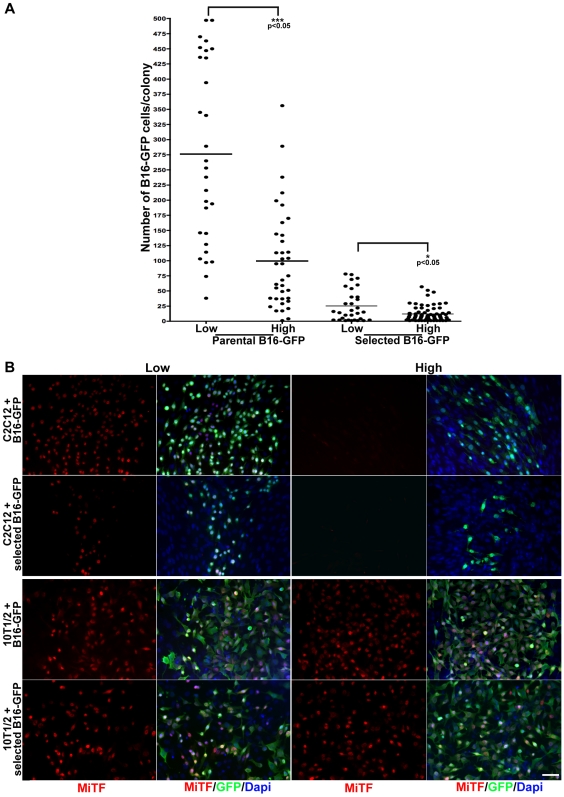
Skeletal muscle is rarely a site of malignant metastasis; the molecular and cellular basis for this rarity is not understood. We report that myogenic cells exert pronounced effects upon co-culture with metastatic melanoma (B16-F10) or carcinoma (LLC1) cells including conversion to the myogenic lineage in vitro and in vivo, as well as inhibition of melanin production in melanoma cells coupled with cytotoxic and cytostatic effects. No effect is seen with non-tumorigenic cells. Tumor suppression assays reveal that the muscle-mediated tumor suppressor effects do not generate resistant clones but function through the down-regulation of the transcription factor MiTF, a master regulator of melanocyte development and a melanoma oncogene. Our findings point to skeletal muscle as a source of therapeutic agents in the treatment of metastatic cancers.


Conflict of interest statement

Competing Interests: The authors have declared that no competing interests exist.

Figures

Figure 1
Figure 1. Myogenic cells inhibit B16-F10 melanoma cells differentiation in vitro.
A) Photomicrographs of GFP expressing melanoma cells (B16) grown alone or in co-culture with myogenic cells (C2C12), fibroblasts (10T1/2), or liver cells (BNL.CL2). We note an inhibition of melanin production in B16 co-cultured with C2C12 as well as a change in their morphology. Scale bar  = 50 µm. B) Quantification of the number of melanin secreting B16 cells grown alone, or in co-culture with fibroblasts, liver cells, or myogenic cells reveals that myogenic cells completely abolish melanin production in B16 cells.
Figure 2
Figure 2. Murine and human muscle cells recruit tumor cells in the myogenic program.
A) Representative photomicrographs of GFP expressing B16 cells (green) grown and differentiated in co-culture with either C2C12 mouse myogenic cell line or mouse primary myoblasts and stained for myosin heavy chain (MF20, red). GFP labeled melanoma cells fuse with the C2C12 cells forming chimeric green myotubes, which are positive for MF20. Nuclei were visualized by DAPI staining (blue). Scale bar  = 20 µm. B) Quantification of GFP expressing myotubes positive for either MyoD or Myosin heavy chain (MF20), from co-cultures of GFP-labeled melanoma cells (B16-GFP) or GFP-labeled Luis Lung Carcinoma cells (LLC1-GFP) with mouse myogenic cells (C2C12), human primary myogenic cells (CHQ), or fibroblasts (10T1/2) immunostained as shown in panel A. Only murine and human muscle cells can recruit Luis Lung Carcinoma (LLC1-GFP) and melanoma(B16-GFP) cells into the myogenic program. This phenotypic conversion is specific to cancer cells and does not occur in co-cultures of GFP labeled fibroblasts with myogenic C2C12 cells. Values represent the mean percentage of at least 3 independent experiments ± SEM. C) Expression of murine Desmin and MyoD transcripts in co-cultures of B16-GFP/CHQ; C2C12/CHQ and CHQ cells alone(RT-PCR). The graph shows the expression levels of Desmin and MyoD transcripts in co-cultures relative to the expression in C2C12/CHQ co-cultures. Experiments were performed in triplicates.
Figure 3
Figure 3. B16 melanoma cells fuse with muscle fibers in vivo.
A-H) Confocal micrographs of representative cross sections of TA muscles, showing fusion of GFP-labeled B16 melanoma cells with injured (A-C) and non-injured (F-H) muscles, 2 weeks after intramuscular injection. The basal lamina is identified by the red laminin immunostaining. Nuclei are visualized by DAPI staining (blue). We note GFP+ myofibers with centrally located nuclei (A-C asterisk), characteristic of muscle fibers undergoing regeneration. Green, GFP+ myofibers are observed in non-regenerating muscle as well, indicating that GFP+ melanoma cells fuse to pre-existing fibers. In both cases, we note GFP positive fibers within the site of injection (A), adjacent to the tumor (F,G), as well as in areas devoid of tumor (B,C,E). At the periphery of the tumor we note a well defined boundary between the tumor and the muscle tissue (arrowheads). No green fibers were detectable in the controlateral control injured and non-injured muscles injected with PBS (D,H). I-K) Representative cross sections of TA muscles, 2 weeks after intrafemoral artery injection of GFP-labeled B16 melanoma cells. L) Confocal micrographs of a representative longitudinal section of TA muscle, showing homogenous expression of GFP along the muscle fiber length, 2 weeks after intramuscular injection of GFP-labeled B16 melanoma cells. Scale bar  = 20 µm.
Figure 4
Figure 4. Apoptotic effect of the conditioned media from C2C12 muscle cells on B16 melanoma cells.
A,B, upper panels) Phase contrast photomicrographs of 10T1/2 fibroblasts (A) and B16-F10 melanoma cells (B) cultured in serum free conditioned media from 10T1/2 fibroblasts (CM10T1/2) or in serum free conditioned media from muscle cells (CMC2C12) for 3 days. After 3 days in CMC2C12, B16 melanoma cells are less numerous and appear rounded and refractive (B). Scale bar  = 50 µm. A,B, lower panels) Propidium iodide flow cytometry cell cycle analysis of 10T1/2 fibroblasts (A) and B16 melanoma cells (B) in the different culture media. The bars indicate the fraction of apoptotic cells for each experimental condition. The cell cycle profile is notably changed in melanoma cells incubated for 3 days in muscle cells conditioned media. C) Percentage of apoptotic versus cycling cells of 10T1/2 fibroblasts (blue bars) and B16-F10 melanoma cells (green bars) cultured in either serum free conditioned media from 10T1/2 fibroblast (CM10T1/2) or C2C12 muscle cells (CMC2C12) after 1 and 3 days post incubation (dpi). Only B16F10 melanoma cells exhibit a significantly high level of apoptotic cells (40.5%±2.5) when cultured in CMC2C12 for 3 days (p≤0.05). Values represent the mean % ± SEM of 3 independent experiments.
Figure 5
Figure 5. Emergence of reversibly resistant B16 melanoma colonies in CMC2C12 during a multiple selection process.
A) Representative photomicrographs of B16 melanoma cells grown in CMC2C12, CM10T1/2, or serum free media (DMEM). CMC2C12 induces pronounced cell death by 3 days. However, 4 days after selection colonies of B16 melanoma cells start to appear. CM10T1/2 and DMEM do not affect the number of melanoma cells. Scale bar  = 40 µm. B) Schematic representation of the number of B16F10 melanoma cells/field after 1, 3 and 4 days of 3 successive rounds of selection in CMC2C12(green), CM10T1/2 (violet) and DMEM (red lines). In the first round of selection (S1) the number of melanoma cells grown in CMC2C12 (green line- circle) sharply decreases at Day 3. Cells grown in CM10T1/2 (violet) or in serum free DMEM (red) continue to proliferate. At 4 day the number of cells grown in CMC2C12 reaches 76% of the original number of cells at time of selection. The same trend is observed for the subsequent rounds of selection (S2,S3). For each time point, values represent a mean of 3 independent experiments. S1: Selection1; S2: Selection2; S3: Selection3. CM10T1/2 (violet lines) (*; p = 0.013). DMEM (red lines) (**; p = 0.006).
Figure 6
Figure 6. Myogenic cells exert density dependant growth inhibition and induce MiTF downregulation on parental and selected B16-GFP melanoma cells.
A) Distribution of the number of cells per colony of either parental or selected B16-GFP melanoma cells, grown with C2C12 cells plated at low and high density. Muscle cells inhibit growth of melanoma cells. Cells previously exposed to muscle conditioned media are more sensitive to this effect. The inhibition is dependent on the density of muscle cell. The black lines represent the mean values of 3 independent experiments: 276 cells/colony for parental B16-GFP in low-density C2C12; 99 cells/colony for parental B16-GFP in high-density C2C12; 25 cells/colony for selected B16-GFP in low-density C2C12; 12 cells/colony for selected B16-GFP in high-density C2C12. B) Left Panels: parental (B16-GFP, green) or selected (selected B16- GFP, green) melanoma cells expressing GFP grown with low density C2C12 or 10T1/2 fibroblasts and immunostained for MiTF (red). MiTF is expressed in parental and selected B16-GFP cells. Fewer B16-GFP cells are present in cultures with C2C12 cells. Nuclei were visualized by Dapi staining (blue). Right panels: B16-GFP and selected B16-GFP melanoma cells grown with high density C2C12 or 10T1/2 fibroblasts and immunostained for MiTF (red). MiTF expression is completely abrogated in parental and selected melanoma cells cultured with C2C12 cells plated at high density. This effect is specific to muscle cells as it is not observed in high density 10T1/2 fibroblasts/B16-GFP co-cultures. Nuclei were counterstained by Dapi (blue). Scale bar  = 50 µm.

Similar articles

Cited by

References

    1. Fidler IJ. Critical determinants of cancer metastasis: rationale for therapy. Cancer Chemother Pharmacol. 1999;43(Suppl):S3–10. - PubMed
    1. Greig RG, Trainer DL. Shaping future strategies for the pharmacological control of tumor cell metastases. Cancer Metastasis Rev. 1986;5:3–14. - PubMed
    1. Klein CA. The direct molecular analysis of metastatic precursor cells in breast cancer: A chance for a better understanding of metastasis and for personalised medicine. Eur J Cancer 2008 - PubMed
    1. Langley RR, Fidler IJ. Tumor cell-organ microenvironment interactions in the pathogenesis of cancer metastasis. Endocr Rev. 2007;28:297–321. - PubMed
    1. Crissman JD. Tumor-host interactions as prognostic factors in the histologic assessment of carcinomas. Pathol Annu. 1986;21 Pt 1:29–52. - PubMed

Publication types

MeSH terms