. 2010 Feb 19;6(2):e1000681.
doi: 10.1371/journal.pcbi.1000681.

Identifying the rules of engagement enabling leukocyte rolling, activation, and adhesion

Affiliations

Identifying the rules of engagement enabling leukocyte rolling, activation, and adhesion

Jonathan Tang et al. PLoS Comput Biol. .

Abstract

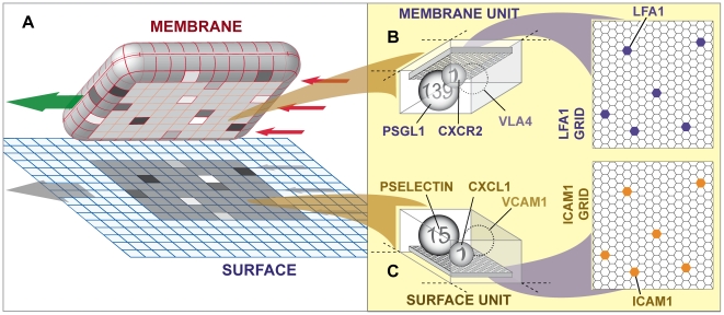
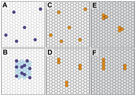
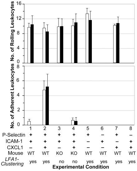
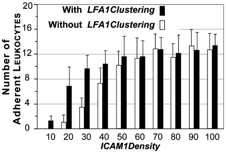
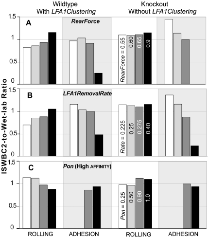
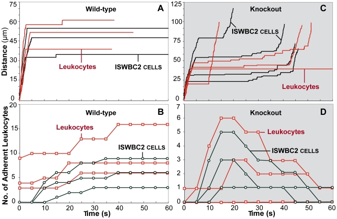
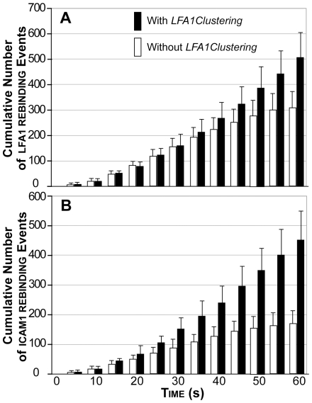
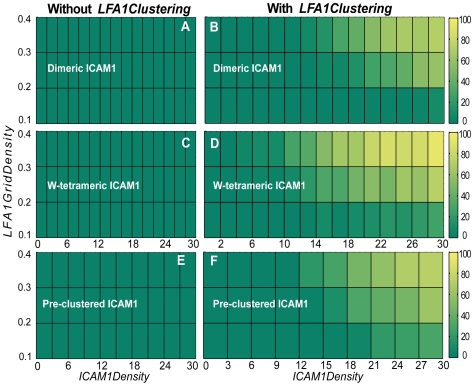
The LFA-1 integrin plays a pivotal role in sustained leukocyte adhesion to the endothelial surface, which is a precondition for leukocyte recruitment into inflammation sites. Strong correlative evidence implicates LFA-1 clustering as being essential for sustained adhesion, and it may also facilitate rebinding events with its ligand ICAM-1. We cannot challenge those hypotheses directly because it is infeasible to measure either process during leukocyte adhesion following rolling. The alternative approach undertaken was to challenge the hypothesized mechanisms by experimenting on validated, working counterparts: simulations in which diffusible, LFA1 objects on the surfaces of quasi-autonomous leukocytes interact with simulated, diffusible, ICAM1 objects on endothelial surfaces during simulated adhesion following rolling. We used object-oriented, agent-based methods to build and execute multi-level, multi-attribute analogues of leukocytes and endothelial surfaces. Validation was achieved across different experimental conditions, in vitro, ex vivo, and in vivo, at both the individual cell and population levels. Because those mechanisms exhibit all of the characteristics of biological mechanisms, they can stand as a concrete, working theory about detailed events occurring at the leukocyte-surface interface during leukocyte rolling and adhesion experiments. We challenged mechanistic hypotheses by conducting experiments in which the consequences of multiple mechanistic events were tracked. We quantified rebinding events between individual components under different conditions, and the role of LFA1 clustering in sustaining leukocyte-surface adhesion and in improving adhesion efficiency. Early during simulations ICAM1 rebinding (to LFA1) but not LFA1 rebinding (to ICAM1) was enhanced by clustering. Later, clustering caused both types of rebinding events to increase. We discovered that clustering was not necessary to achieve adhesion as long as LFA1 and ICAM1 object densities were above a critical level. Importantly, at low densities LFA1 clustering enabled improved efficiency: adhesion exhibited measurable, cell level positive cooperativity.


Conflict of interest statement

The authors have declared that no competing interests exist.

Figures

Figure 1
Figure 1. Sketch of the ISWBC2 experimental system components.
(A) A leukocyte agent is shown pulled away from the simulated surface to which it was attached. The left arrow indicates roll direction; the three right arrows indicate shear resulting from the simulated flow. The surface is discretized into independent units of function called surface units. The leukocyte's membrane is similarly discretized into matching units called membrane units: 600 total (20×30). The 8×10 shaded region on the surface and on the leukocyte's underside identifies the contact zone. The units within the contact zones that are shaded differently indicate that different numbers of bonds had formed between ligandligand pairs in overlapping units; otherwise, no bonds formed. Rolling is the result of a sequence of forward ratchet events. One ratchet event is the result of one row of membrane units being released at the rear of the contact zone along with engagement of a new row of at the front of the contact zone. One ratchet event maps to a leukocyte rolling approximately 1 µm (relative to the flow chamber surface). (B) A membrane unit is illustrated. Each is a software object functioning as a container. All leukocyte membrane functionality (relevant to these studies) within each unit of surface on icam1Grid is represented by the four objects: psgl1, vla4 (not used for most experiments), cxcr2, and lfa1. They map to leukocyte receptors; they are illustrated as spheres. The number on each sphere indicates the number of receptors to which that agent maps. Mobile lfa1 objects reside on a lfa1Grid. Each lfa1 maps to an individual LFA-1 molecule. (C) A surface unit is illustrated. Similar to membrane units, each surface unit is simulated using a software object functioning as a container. All flow chamber surface functionality (relevant to these studies) within each surface unit is represented by four objects: pselectin, vcam1 (not used for most experiments), cxcl1, and icam1. They map to surface receptors and chemokines. The number on each sphere indicates the number of receptors or chemokines to which that agent maps. Similar to lfa1, an icam1 resides on icam1Grid, a 2D hexagonal lattice, and maps to an individual ICAM-1 molecule.
Figure 2
Figure 2. Diagrams illustrate the different spatial configurations of lfa1 and icam1 adhesion molecules implemented and tested during simulations.
lfa1Grids are shown with lfa1 objects labeled dark blue in either (A) a nonclustered state or (B) a clustered state. Clustered lfa1 are spatially restricted within the light blue region. Icam1Grids are shown with icam1 objects labeled yellow in either a (C) native monomeric state, (D) a native dimeric state, (E) as preformed clusters, or (F) as w-tetramers formed after ligand-binding.
Figure 3
Figure 3. Ex vivo and in silico results for eight different experimental conditions are compared.
Ex vivo conditions (from [13]): the flow chamber surface was coated with P-selectin and/or ICAM-1 with or without immobilized CXCL1 chemokine. Mice were either WT or PI3Kγ knockouts (KO). Leukocytes that rolled and adhered within each of five fields of view were recorded during a 60-second observation interval. White bars are ex vivo means ± 1 SE. The paired black bars are ISWBC2 means ± 1 SD (20 populations containing 30 leukocytes each) for the same condition using the parameter values in Tables 4 and 5. ISWBC2 experiments that map to WT mouse counterparts used LFA1Clustering = true. Simulations of KO mice used the parameter setting LFA1Clustering = false.
Figure 4
Figure 4. Effect of varying ICAM1Density parameter values on leukocyte adhesion.
The effect of Leukocyte ICAM1Density was varied from 10 to 100 at intervals of 10. Bar heights are ISWBC2 means ± 1 SD (20 populations containing 30 leukocytes each) for the same condition using the parameter values in Tables 4 and 5. Black bars indicate simulation experiments when LFA1Clustering = true. White bars indicate simulation experiments when LFA1Clustering = false.
Figure 5
Figure 5. Robustness of ISWBC2s to changes in the LFA1RemovalRate, Pon, and RearForce.
Separate sets of in silico experiments, using the leukocyte parameter values in Table 4 and environment parameter values in Table 5, were completed while varying either (A) RearForce, (B) LFA1RemovalRate, or (C) Pon(high affinity lfa1-icam1) as indicated. Bars heights are ratios of ISWBC2-to-wet-lab results of rolling and adhesion data for both WT and KO mice of the type used for the experiments in Figure 3. Comparable adjustments of other parameters caused similar gradual changes in leukocyte rolling and adhesion data. The listed parameter values for rolling under Knockout also apply to the similarly shaded bar for the other three conditions.
Figure 6
Figure 6. In vivo and in silico results are compared.
In vivo conditions (from [13]): mice were either (A, B) WT or (C, D) KO. Red: wet-lab values; black: ISWBC2 values. Wet-lab experiments: post-capillary venules were observed from one minute before to one minute after CXCL1 injection at t = 0. Each simulation ran for 600 simulation cycles (equivalent to about 60 seconds). Individual leukocyte trajectories are plotted for (A) WT and (C) KO mice. (B, D) Individual leukocytes were tracked every 5 s, and those that were adherent during that time were counted. Open squares are adherent leukocyte counts for individual venules. Open circles are corresponding adherent leukocyte counts for venules. Thirty leukocytes comprised the population within a venule. Individual leukocytes were tracked every 50 simulation cycles (approximately 5 s), and those that were adherent were counted. ISWBC2: parameter values are those listed in Tables 4 and 5, and leukocytes are in the presence of cxcl1 beginning at t = 0. Lfa1 clustering was either (A, B) enabled or (C, D) disabled.
Figure 7
Figure 7. Effect of lfa clustering on lfa1 and icam1 re-binding events.
The cumulative number of (A) lfa1 re-binding events and (B) icam1 re-binding events were counted at 50 simulation cycle intervals (5 seconds) for each leukocyte. A lfa1 (or icam1) rebinding event was defined as a bond formation event by a lfa1 (or icam1) object that had already participated in a bond formation event in a previous time step. Averages are plotted for 30 leukocytes per experimental condition (with or without LFA1Clustering). Simulation data with LFA1Clustering were from leukocytes that sustained adhesion (rolling followed by at least 30 simulation cycles of arrest until the end of the simulation). Simulation data without LFA1Clustering were from leukocytes that exhibited initial and transient adhesion (at least 300 simulation cycles of arrest). Without LFA1Clustering, the average adhesion time was 39.8±7.2 seconds before the leukocyte initiated rolling again (average time = 43.9±7.1 seconds). Error bars: ± SD.
Figure 8
Figure 8. The effect on sustained adhesion of different hypothesized icam1 configurations.
The percentage of leukocytes that sustained adhesion (color scale at right) were calculated for varying LFA1GridDensity and ICAM1Density values using the different hypothesized icam1 configurations discussed in the text. Lfa1 clustering was either disabled (left: A, C, E) or enabled (right: B, D, F). Icam1 was (A, B) dimeric, (C, D) dimeric and allowed to form linear tetramers upon ligand-binding, or (E, F) was preformed into nanoclusters. No sustained adhesion occurred when icam1 was monomeric (not shown).

Similar articles

Cited by

References

    1. Kinashi T. Intracellular Signalling Controlling Integrin Activation in Lymphocytes. Nat Rev Immunol. 2005;5:546–59. - PubMed
    1. Kim M, Carman CV, Yang W, Salas A, Springer TA. The primacy of affinity over clustering in regulation of adhesiveness of the integrin {alpha}L{beta}2. J Cell Biol. 2004;167:1241–1253. - PMC - PubMed
    1. Lum AF, Green CE, Lee GR, Staunton DE, Simon SI. Dynamic regulation of LFA-1 activation and neutrophil arrest on intercellular adhesion molecule 1 (ICAM-1) in shear flow. J Biol Chem. 2002;277:20660–70. - PubMed
    1. Constantin G, Majeed M, Giagulli C, Piccio L, Kim JY, et al. Chemokines trigger immediate beta2 integrin affinity and mobility changes: differential regulation and roles in lymphocyte arrest under flow. Immunity. 2000;13:759–769. - PubMed
    1. Sarantos MR, Raychaudhuri S, Lum AFH, Staunton DE, Simon SI. Leukocyte function-associated antigen 1-mediated adhesion stability is dynamically regulated through affinity and valency during bond formation with intercellular adhesion molecule-1. J Biol Chem. 2005;280:28290–28298. - PubMed

Publication types

Substances