. 2010 Feb 19;6(2):e1000772.
doi: 10.1371/journal.ppat.1000772.

Arterivirus Nsp1 modulates the accumulation of minus-strand templates to control the relative abundance of viral mRNAs

Affiliations

Arterivirus Nsp1 modulates the accumulation of minus-strand templates to control the relative abundance of viral mRNAs

Danny D Nedialkova et al. PLoS Pathog. .

Abstract

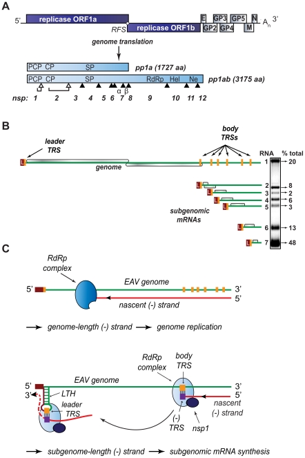
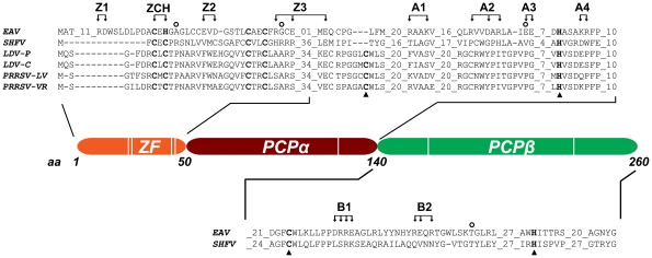
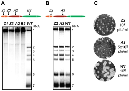
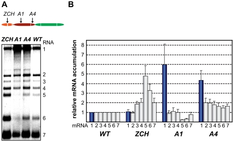
The gene expression of plus-strand RNA viruses with a polycistronic genome depends on translation and replication of the genomic mRNA, as well as synthesis of subgenomic (sg) mRNAs. Arteriviruses and coronaviruses, distantly related members of the nidovirus order, employ a unique mechanism of discontinuous minus-strand RNA synthesis to generate subgenome-length templates for the synthesis of a nested set of sg mRNAs. Non-structural protein 1 (nsp1) of the arterivirus equine arteritis virus (EAV), a multifunctional regulator of viral RNA synthesis and virion biogenesis, was previously implicated in controlling the balance between genome replication and sg mRNA synthesis. Here, we employed reverse and forward genetics to gain insight into the multiple regulatory roles of nsp1. Our analysis revealed that the relative abundance of viral mRNAs is tightly controlled by an intricate network of interactions involving all nsp1 subdomains. Distinct nsp1 mutations affected the quantitative balance among viral mRNA species, and our data implicate nsp1 in controlling the accumulation of full-length and subgenome-length minus-strand templates for viral mRNA synthesis. The moderate differential changes in viral mRNA abundance of nsp1 mutants resulted in similarly altered viral protein levels, but progeny virus yields were greatly reduced. Pseudorevertant analysis provided compelling genetic evidence that balanced EAV mRNA accumulation is critical for efficient virus production. This first report on protein-mediated, mRNA-specific control of nidovirus RNA synthesis reveals the existence of an integral control mechanism to fine-tune replication, sg mRNA synthesis, and virus production, and establishes a major role for nsp1 in coordinating the arterivirus replicative cycle.


Conflict of interest statement

The authors have declared that no competing interests exist.

Figures

Figure 1
Figure 1. Organization and expression of the polycistronic EAV +RNA genome.
(A) Top: EAV genome organization, showing the 5′-proximal replicase open reading frames (ORFs), as well as the downstream ORFs encoding the viral structural proteins envelope (E), membrane (M), nucleocapsid (N), and glycoproteins (GP) 2–5 and the 3′ poly(A) tail (An). Bottom: overview of the pp1a and pp1ab replicase polyproteins that result from genome translation, which requires an ORF1a/1b ribosomal frameshift (RFS) to produce pp1ab. Arrowheads represent sites cleaved by the three virus-encoded proteases (open for autoproteolytically processed ones, closed for sites processed by the main proteinase in nsp4). The resulting nonstructural proteins (nsp) are numbered. The key viral enzymatic domains such as the nsp1 papain-like cysteine proteinase β (PCP), nsp2 cysteine proteinase (CP), nsp4 serine proteinase (SP), nsp9 viral RNA-dependent RNA polymerase (RdRp), nsp10 helicase (Hel), and nsp11 endoribonuclease (Ne) are indicated. (B) Overview of viral mRNA species produced in EAV-infected cells. The ORFs expressed from the respective mRNAs are shown in gray, and the 5′ leader sequence is depicted in dark red. The orange boxes indicate the positions of transcription-regulating sequences (TRS). The gel hybridization image on the right is representative of the wild-type accumulation levels of the seven EAV mRNAs at the time point used for analysis in the study (see text for details). The amount of each mRNA, determined by quantitative phosphorimager analysis, is indicated as percentage of the total amount of viral mRNA. (C) Model for EAV replication and transcription. Continuous minus-strand RNA synthesis yields a genome-length minus strand template for genome replication, a process for which nsp1 is dispensable. Discontinuous minus-strand RNA synthesis results in a nested set of subgenome-length minus strands that serve as templates for sg mRNA synthesis (see text for details). Nsp1 is crucial for this process, which is also guided by a base pairing interaction between the TRS complement [(−)TRS] at the 3′ end of the nascent minus-strand and the genomic leader TRS, present in a RNA hairpin structure (LTH).
Figure 2
Figure 2. Domain organization of EAV nsp1.
The partial sequence alignment shows key regions in the three subdomains previously identified in the arterivirus nsp1 region. GenBank accession numbers for the full-length arterivirus genomes used for the alignment are as follows: EAV, NC_002532; simian hemorrhagic fever virus (SHFV), NC_003092; lactate dehydrogenase-elevating virus (LDV-P and LDV-C), NC_001639 and NC_002534; PRRSV-LV, M96262.2; PRRSV-VR, AY150564. Zinc-coordinating residues are indicated in bold font; the active-site Cys and His of PCPα and PCPβ are indicated with triangles (note the loss of the active-site Cys in EAV PCPα). The positions of amino acid clusters mutated in this study are indicated with arrows. All substitutions were with Ala, with the exception of the ZCH construct, in which Cys-25 and His-27 were swapped. The positions of mutations found in pseudorevertants are indicated with open circles.
Figure 3
Figure 3. Importance of nsp1 subdomains for transcription and virus production.
(A, B). Analysis of EAV-specific mRNA accumulation by gel hybridization. The domain organization of nsp1 is depicted as in Fig. 2 and the positions of the clusters of amino acid mutations analyzed are indicated with arrows. BHK-21 cells were transfected with RNA transcribed from wt or selected mutant EAV full-length cDNA clones. Total intracellular RNA was isolated at 11 h post-transfection and resolved by denaturing formaldehyde electrophoresis. Equal loading of samples was confirmed by ethidium bromide staining of ribosomal RNA (data not shown). EAV-specific mRNAs were detected by hybridization of the gel with a 32P-labelled probe complementary to the 3′-end of the viral genome and subsequent phosphorimaging. The positions of the EAV genome (RNA1) and the six sg mRNAs (RNA2 to RNA7) are indicated. (C) Plaque phenotype and virus titers of the Z2 and A3 mutants. Plaque assays were performed on BHK-21 using cell culture supernatants harvested 24 h after transfection. Cells were incubated under a semi-solid overlay at 39.5°C for 72 h, fixed and stained with crystal violet. Virus titers represent an average of three independent experiments. Pfu, plaque-forming units.
Figure 4
Figure 4. Multiple mutations in nsp1 exert species-specific effects on viral mRNA accumulation.
(A, B) Gel hybridization analysis and quantification of EAV-specific mRNA accumulation in cells transfected with the ZCH, A1, A4 mutant or a wt control. (A) Viral mRNA accumulation was analyzed at 11 h post-transfection by gel hybridization as described in the legend to Fig. 3. (B) The accumulation levels of each viral mRNA in the nsp1 mutants were quantified by phosphorimaging in the linear range of exposure and normalized to the level of accumulation of each corresponding viral mRNA in the wt control, which was set at 1. Genomic RNA levels are represented as blue bars. The relative values correspond to the means from three independent transfections and error bars denote standard deviation.
Figure 5
Figure 5. Minus-strand RNA accumulation is also modulated by mutations in nsp1.
(A–D) Analysis and quantification of EAV minus-strand accumulation by a two-cycle RNase protection assay. (A) Schematic representation of the nested set of viral minus-strand RNA [(−)RNA] species produced in EAV-infected cells. The anti-leader sequence is depicted in light green. The in vitro-transcribed plus-strand probes used for detection of (−)RNA1 (pRNA1), (−)RNA6 (pRNA6) and (−) RNA7 (pRNA7) are shown. pRNA6 and pRNA7 target the leader-body junction sequences of (−)RNA6 and (−)RNA7, respectively. Note that hybridization with pRNA1 results in the protection of a single fragment, while the probes for (−)RNAs 6 and 7 each protect three fragments – one derived from the full-length sg minus strand, and two fragments derived in part from partial hybridization of these probes to larger viral (−)RNAs in which the target sequences are noncontiguous (exemplified for pRNA6). For simplicity, non-EAV sequences present near the termini of the three probes were omitted from the scheme. (B) Viral (−)RNA accumulation was analyzed at 11 h post-transfection for the ZCH, A1 and A4 mutants, and a wt control. Protected fragments were resolved on denaturing 5% polyacrylamide/8M urea gels and visualized by phosphorimaging. The constructs analyzed are labeled above the lanes (M, mock-transfected cells; (−), no-RNase control that shows a band corresponding to 0.2 fmol of the full-length probe). Sizes (nt) of RNA markers have been indicated on the left. The single 327-nt protected fragment resulting from hybridization with the positive-sense probe for RNA1(−) is indicated. The probes for subgenome-length minus strands protected fragments derived from the full-length (−)RNA6 and (−)RNA7 (327 nt and 319 nt, respectively; denoted with LB), as well as from the (−)RNA6 and (−)RNA7 body sequences (188 nt and 180 nt, respectively; denoted with B) and the anti-leader sequence (139 nt; denoted with L). The presence of two bands in the size range of the anti-leader fragment has been described previously . (C) The relative levels of minus-strand accumulation were quantified by phosphorimaging. For (−)RNAs 6 and 7, only the bands resulting from protection of full-length sg minus strands (denoted with LB in panel [B]) were quantified. The values correspond to the means from three independent transfections that were normalized to the level of accumulation of each minus-strand RNA in the wt control, which was set at 1. Intracellular RNA from the same transfection samples for which plus-strand accumulation was quantified (Fig. 4B) was used. Genomic minus-strand RNA levels are represented as dark blue bars. Error bars denote standard deviation. (D) The ratio of plus-strand to minus-strand accumulation for RNAs 1, 6 and 7 was calculated using the mean relative values obtained in Fig. 4B and Fig. 5C.
Figure 6
Figure 6. Analysis of viral protein accumulation and virus production by selected nsp1 mutants.
(A) Western blot analysis of EAV-specific protein accumulation. Cells transfected with wt or mutant viral genomes were harvested 11 h after transfection and equal amounts of total protein were analyzed with EAV-specific sera detecting nsp3, M and N, which are translated from RNAs 1, 6 and 7, respectively. The relative levels of each mRNA template (derived from Fig. 4B) are indicated below the gels. Beta-actin was used as a loading control. (B) Plaque phenotype and virus titers of the ZCH, A1 and A4 mutants in comparison with wt. Plaque assays were performed on BHK-21 using cell culture supernatants harvested 24 h after transfection. Virus titers represent an average of three independent experiments.
Figure 7
Figure 7. Second-site mutations in nsp1 moderate species-specific defects in mRNA accumulation.
(A–D) Gel hybridization analysis and quantification of EAV-specific mRNA accumulation. (A) BHK-21 cells were transfected with ZCH and A1 mutants, reconstructed pseudorevertants, and wt controls. The positions of the originally mutated amino acid clusters are indicated with arrows; the open circle denotes the position of second-site mutations. Viral mRNAs were analyzed 11 h post-transfection by gel hybridization as described above. (B) For ZCH+A29D and A1+A29K, the accumulation levels of each viral mRNA were quantified by phosphorimaging and the values were normalized to the wt level of accumulation of each corresponding viral mRNA from the same experiment, set at 1. Genomic RNA levels are shown as blue bars. The relative values correspond to the means from three independent transfections and error bars denote the standard deviation. The relative accumulation levels of viral mRNAs at 11 h post-transfection for the ZCH and A1 mutants are derived from Fig. 4B and are represented here to facilitate comparison between mutant and pseudorevertant phenotypes. (C) BHK-21 cells were transfected with the A4 mutant, reconstructed pseudorevertants and a wt control. The positions of the originally mutated amino acid cluster and the second-site mutations are indicated as in (A). Viral mRNAs were analyzed 11 h post-transfection. (D) For A4+G47A, A4+E112K, and A4+T196K, quantification of relative viral mRNA accumulation levels was performed as described in (B). Similarly, the relative accumulation levels of viral mRNAs at 11 h post-transfection for the A4 mutant derived from Fig. 4B are represented to facilitate comparison.
Figure 8
Figure 8. Relationship between viral mRNA accumulation profiles and infectious virus yield.
The data obtained on the accumulation of genome and sg mRNAs were used for a quantitative assessment of nsp1 mutant phenotypes in terms of (A) changes in mRNA accumulation compared to wt and (B) the extent to which the balance between viral transcripts was disturbed (B). (A) For each mutant and pseudorevertant, a value for the accumulation of each of its 7 mRNAs at 11 h post-transfection had been assigned as compared to that of the wt control (see Fig. 4B, 7B and 7D). The mean of these seven values (“mean relative mRNA accumulation”) was plotted against the corresponding progeny virus titer in culture supernatants at 24 h post-transfection (Fig. 6B and table 2). Wild-type is depicted in orange. The engineered nsp1 mutants and nsp1 pseudorevertants are shown in blue and purple, respectively. The three pseudorevertants of mutant A4, displaying very similar phenotypes, are indicated as A4+PSR. (B) For each mRNA species, the deviation of its relative accumulation from the mean of the complete nested set of mRNAs (see panel A) was calculated. From these seven values, the mean (absolute) deviation was calculated for each mutant and plotted against virus titers as in (A). For wt, the mean deviation is 0. Engineered nsp1 mutants and their pseudorevertants are indicated as in (A). The data fit a negative exponential regression calculated using Microsoft Excel and depicted as a gray line (y = 6×10⁶+7e−8.4306x, R2 = 0.95). The inset shows an expanded view of the upper left quadrant of the graph (shaded in gray).

Similar articles

Cited by

References

    1. Miller WA, Koev G. Synthesis of subgenomic RNAs by positive-strand RNA viruses. Virology. 2000;273:1–8. - PubMed
    1. Perlman S, Netland J. Coronaviruses post-SARS: update on replication and pathogenesis. Nat Rev Microbiol. 2009;7:439–450. - PMC - PubMed
    1. Gorbalenya AE, Enjuanes L, Ziebuhr J, Snijder EJ. Nidovirales: evolving the largest RNA virus genome. Virus Res. 2006;117:17–37. - PMC - PubMed
    1. Molenkamp R, van Tol H, Rozier BCD, van der Meer Y, Spaan WJM, Snijder EJ. The arterivirus replicase is the only viral protein required for genome replication and subgenomic mRNA transcription. J Gen Virol. 2000;81:2491–2496. - PubMed
    1. Sawicki SG, Sawicki DL. Coronaviruses use discontinuous extension for synthesis of subgenome-length negative strands. Adv Exp Med Biol. 1995;380:499–506. - PubMed

Publication types