

Neutrophils are resistant to Yersinia YopJ/P-induced apoptosis and are protected from ROS-mediated cell death by the type III secretion system
- PMID: 20174624
- PMCID: PMC2823771
- DOI: 10.1371/journal.pone.0009279
Neutrophils are resistant to Yersinia YopJ/P-induced apoptosis and are protected from ROS-mediated cell death by the type III secretion system
Abstract
Background: The human innate immune system relies on the coordinated activity of macrophages and polymorphonuclear leukocytes (neutrophils or PMNs) for defense against bacterial pathogens. Yersinia spp. subvert the innate immune response to cause disease in humans. In particular, the Yersinia outer protein YopJ (Y. pestis and Y. pseudotuberculosis) and YopP (Y. enterocolitica) rapidly induce apoptosis in murine macrophages and dendritic cells. However, the effects of Yersinia Yop J/P on neutrophil fate are not clearly defined.
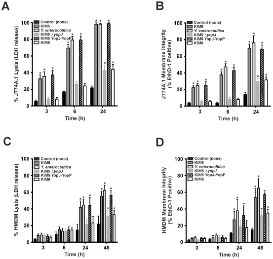
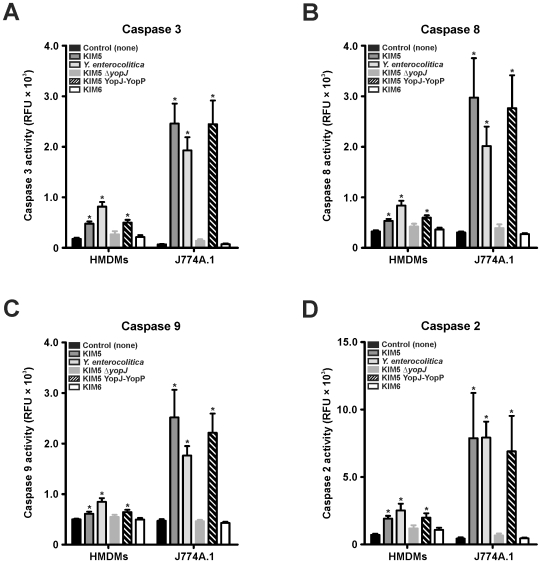
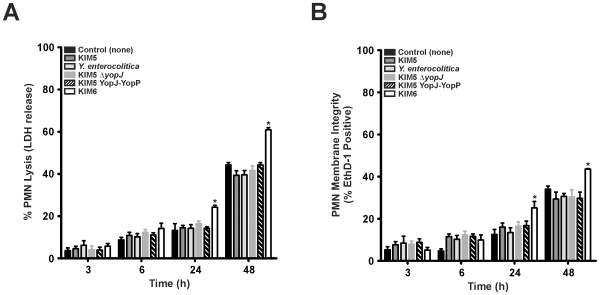
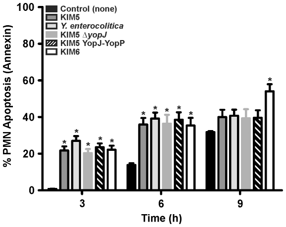
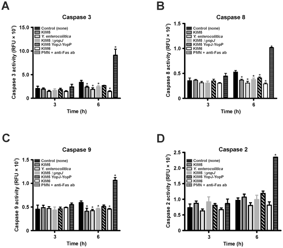
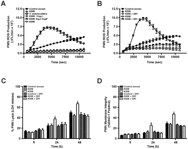
Methodology/principal findings: In this study, we utilized wild-type and mutant strains of Yersinia to test the contribution of YopJ and YopP on induction of apoptosis in human monocyte-derived macrophages (HMDM) and neutrophils. Whereas YopJ and YopP similarly induced apoptosis in HMDMs, interaction of human neutrophils with virulence plasmid-containing Yersinia did not result in PMN caspase activation, release of LDH, or loss of membrane integrity greater than PMN controls. In contrast, interaction of human PMNs with the virulence plasmid-deficient Y. pestis strain KIM6 resulted in increased surface exposure of phosphatidylserine (PS) and cell death. PMN reactive oxygen species (ROS) production was inhibited in a virulence plasmid-dependent but YopJ/YopP-independent manner. Following phagocytic interaction with Y. pestis strain KIM6, inhibition of PMN ROS production with diphenyleneiodonium chloride resulted in a reduction of PMN cell death similar to that induced by the virulence plasmid-containing strain Y. pestis KIM5.
Conclusions: Our findings showed that Yersinia YopJ and/or YopP did not induce pronounced apoptosis in human neutrophils. Furthermore, robust PMN ROS production in response to virulence plasmid-deficient Yersinia was associated with increased PMN cell death, suggesting that Yersinia inhibition of PMN ROS production plays a role in evasion of the human innate immune response in part by limiting PMN apoptosis.
Conflict of interest statement
Figures
Similar articles
-
YopJ-induced caspase-1 activation in Yersinia-infected macrophages: independent of apoptosis, linked to necrosis, dispensable for innate host defense.PLoS One. 2012;7(4):e36019. doi: 10.1371/journal.pone.0036019. Epub 2012 Apr 26. PLoS One. 2012. PMID: 22563435 Free PMC article.
-
Reduced secretion of YopJ by Yersinia limits in vivo cell death but enhances bacterial virulence.PLoS Pathog. 2008 May 16;4(5):e1000067. doi: 10.1371/journal.ppat.1000067. PLoS Pathog. 2008. PMID: 18483548 Free PMC article.
-
Interaction of Yersinia pestis with macrophages: limitations in YopJ-dependent apoptosis.Infect Immun. 2006 Jun;74(6):3239-50. doi: 10.1128/IAI.00097-06. Infect Immun. 2006. PMID: 16714551 Free PMC article.
-
The virulence plasmid of Yersinia, an antihost genome.Microbiol Mol Biol Rev. 1998 Dec;62(4):1315-52. doi: 10.1128/MMBR.62.4.1315-1352.1998. Microbiol Mol Biol Rev. 1998. PMID: 9841674 Free PMC article. Review.
-
Yersinia type III effectors perturb host innate immune responses.World J Biol Chem. 2016 Feb 26;7(1):1-13. doi: 10.4331/wjbc.v7.i1.1. World J Biol Chem. 2016. PMID: 26981193 Free PMC article. Review.
Cited by
-
Disruption of fas-fas ligand signaling, apoptosis, and innate immunity by bacterial pathogens.PLoS Pathog. 2014 Aug 7;10(8):e1004252. doi: 10.1371/journal.ppat.1004252. eCollection 2014 Aug. PLoS Pathog. 2014. PMID: 25101900 Free PMC article. Review. No abstract available.
-
Non-canonical autophosphorylation of RIPK1 drives timely pyroptosis to control Yersinia infection.Cell Rep. 2024 Aug 27;43(8):114641. doi: 10.1016/j.celrep.2024.114641. Epub 2024 Aug 17. Cell Rep. 2024. PMID: 39154339 Free PMC article.
-
Inhibition of Neutrophil Primary Granule Release during Yersinia pestis Pulmonary Infection.mBio. 2019 Dec 10;10(6):e02759-19. doi: 10.1128/mBio.02759-19. mBio. 2019. PMID: 31822588 Free PMC article.
-
RIPK1 activates distinct gasdermins in macrophages and neutrophils upon pathogen blockade of innate immune signaling.Proc Natl Acad Sci U S A. 2021 Jul 13;118(28):e2101189118. doi: 10.1073/pnas.2101189118. Proc Natl Acad Sci U S A. 2021. PMID: 34260403 Free PMC article.
-
Yersinia pestis Actively Inhibits the Production of Extracellular Vesicles by Human Neutrophils.J Extracell Vesicles. 2025 Apr;14(4):e70074. doi: 10.1002/jev2.70074. J Extracell Vesicles. 2025. PMID: 40240908 Free PMC article.
References
-
- Hume DA. The mononuclear phagocyte system. Curr Opin Immunol. 2006;18:49–53. - PubMed
-
- Nauseef WM, Clark RA. Granulocytic Phagocytes. In: Mandel GL, Bennett JE, Dolin R, editors. Basic principles in the diagnosis and management of infectious diseases. New York: Churchill Livingstone; 2005. pp. 93–117.
-
- DeLeo F. Modulation of phagocyte apoptosis by bacterial pathogens. Apoptosis. 2004;9:399–413. - PubMed
-
- Kobayashi SD, Voyich JM, Braughton KR, Whitney AR, Nauseef WN, et al. Gene expression profiling provides insight into the pathophysiology of chronic granulomatous disease. J Immunol. 2004;172:636–643. - PubMed
-
- Sanmun D, Witasp E, Jitkaew S, Tyurina YY, Kagan VE, et al. Involvement of a functional NADPH oxidase in neutrophils and macrophages during programmed cell clearance: implications for chronic granulomatous disease. Am J Physiol Cell Physiol. 2009;3:C621–631. - PubMed
Publication types
MeSH terms
Substances
Associated data
- Actions
- Actions
Grants and funding
LinkOut - more resources
Full Text Sources
Other Literature Sources
Miscellaneous
