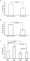
ZNF9 activation of IRES-mediated translation of the human ODC mRNA is decreased in myotonic dystrophy type 2
- PMID: 20174632
- PMCID: PMC2823779
- DOI: 10.1371/journal.pone.0009301
ZNF9 activation of IRES-mediated translation of the human ODC mRNA is decreased in myotonic dystrophy type 2
Abstract
Myotonic dystrophy types 1 and 2 (DM1 and DM2) are forms of muscular dystrophy that share similar clinical and molecular manifestations, such as myotonia, muscle weakness, cardiac anomalies, cataracts, and the presence of defined RNA-containing foci in muscle nuclei. DM2 is caused by an expansion of the tetranucleotide CCTG repeat within the first intron of ZNF9, although the mechanism by which the expanded nucleotide repeat causes the debilitating symptoms of DM2 is unclear. Conflicting studies have led to two models for the mechanisms leading to the problems associated with DM2. First, a gain-of-function disease model hypothesizes that the repeat expansions in the transcribed RNA do not directly affect ZNF9 function. Instead repeat-containing RNAs are thought to sequester proteins in the nucleus, causing misregulation of normal cellular processes. In the alternative model, the repeat expansions impair ZNF9 function and lead to a decrease in the level of translation. Here we examine the normal in vivo function of ZNF9. We report that ZNF9 associates with actively translating ribosomes and functions as an activator of cap-independent translation of the human ODC mRNA. This activity is mediated by direct binding of ZNF9 to the internal ribosome entry site sequence (IRES) within the 5'UTR of ODC mRNA. ZNF9 can activate IRES-mediated translation of ODC within primary human myoblasts, and this activity is reduced in myoblasts derived from a DM2 patient. These data identify ZNF9 as a regulator of cap-independent translation and indicate that ZNF9 activity may contribute mechanistically to the myotonic dystrophy type 2 phenotype.
Conflict of interest statement
Figures


Similar articles
-
The myotonic dystrophy type 2 protein ZNF9 is part of an ITAF complex that promotes cap-independent translation.Mol Cell Proteomics. 2007 Jun;6(6):1049-58. doi: 10.1074/mcp.M600384-MCP200. Epub 2007 Feb 26. Mol Cell Proteomics. 2007. PMID: 17327219
-
Reduction of the rate of protein translation in patients with myotonic dystrophy 2.J Neurosci. 2009 Jul 15;29(28):9042-9. doi: 10.1523/JNEUROSCI.1983-09.2009. J Neurosci. 2009. PMID: 19605641 Free PMC article.
-
CCUG repeats reduce the rate of global protein synthesis in myotonic dystrophy type 2.Rev Neurosci. 2010;21(1):19-28. doi: 10.1515/revneuro.2010.21.1.19. Rev Neurosci. 2010. PMID: 20458885 Review.
-
Effect of the [CCTG]n repeat expansion on ZNF9 expression in myotonic dystrophy type II (DM2).Biochim Biophys Acta. 2006 Mar;1762(3):329-34. doi: 10.1016/j.bbadis.2005.11.004. Epub 2005 Dec 6. Biochim Biophys Acta. 2006. PMID: 16376058
-
Myotonic dystrophies: An update on clinical aspects, genetic, pathology, and molecular pathomechanisms.Biochim Biophys Acta. 2015 Apr;1852(4):594-606. doi: 10.1016/j.bbadis.2014.05.019. Epub 2014 May 29. Biochim Biophys Acta. 2015. PMID: 24882752 Review.
Cited by
-
Alternative Mechanisms to Initiate Translation in Eukaryotic mRNAs.Comp Funct Genomics. 2012;2012:391546. doi: 10.1155/2012/391546. Epub 2012 Feb 16. Comp Funct Genomics. 2012. PMID: 22536116 Free PMC article.
-
Phosphomimetic substitution of heterogeneous nuclear ribonucleoprotein A1 at serine 199 abolishes AKT-dependent internal ribosome entry site-transacting factor (ITAF) function via effects on strand annealing and results in mammalian target of rapamycin complex 1 (mTORC1) inhibitor sensitivity.J Biol Chem. 2011 May 6;286(18):16402-13. doi: 10.1074/jbc.M110.205096. Epub 2011 Mar 16. J Biol Chem. 2011. PMID: 21454539 Free PMC article.
-
Bridging integrator 1 (BIN1) protein expression increases in the Alzheimer's disease brain and correlates with neurofibrillary tangle pathology.J Alzheimers Dis. 2014;42(4):1221-7. doi: 10.3233/JAD-132450. J Alzheimers Dis. 2014. PMID: 25024306 Free PMC article.
-
Yeast Gis2 and its human ortholog CNBP are novel components of stress-induced RNP granules.PLoS One. 2012;7(12):e52824. doi: 10.1371/journal.pone.0052824. Epub 2012 Dec 21. PLoS One. 2012. PMID: 23285195 Free PMC article.
-
The role of CNBP in brain atrophy and its targeting in myotonic dystrophy type 2.Hum Mol Genet. 2025 Mar 7;34(6):512-522. doi: 10.1093/hmg/ddaf002. Hum Mol Genet. 2025. PMID: 39807631
References
-
- Cho DH, Tapscott SJ. Myotonic dystrophy: emerging mechanisms for DM1 and DM2. Biochimica et Biophysica Acta. 2007;1772:195–204. - PubMed
-
- Aslanidis C, Jansen G, Amemiya C, Shutler G, Mahadevan M, et al. Cloning of the essential myotonic dystrophy region and mapping of the putative defect. Nature. 1992;355:548–551. - PubMed
-
- Liquori CL, Ricker K, Moseley ML, Jacobsen JF, Kress W, et al. Myotonic dystrophy type 2 caused by a CCTG expansion in intron 1 of ZNF9. Science. 2001;293:864–867. - PubMed
-
- Ranum LP, Day JW. Myotonic dystrophy: clinical and molecular parallels between myotonic dystrophy type 1 and type 2. Curr Neurol Neurosci Rep. 2002;2:465–470. - PubMed
-
- Day JW, Ricker K, Jacobsen JF, Rasmussen LJ, Dick KA, et al. Myotonic dystrophy type 2: molecular, diagnostic and clinical spectrum. Neurology. 2003;60:657–664. - PubMed
Publication types
MeSH terms
Substances
Grants and funding
LinkOut - more resources
Full Text Sources
Research Materials
Miscellaneous

