

Enriched population of PNS neurons derived from human embryonic stem cells as a platform for studying peripheral neuropathies
- PMID: 20174633
- PMCID: PMC2823780
- DOI: 10.1371/journal.pone.0009290
Enriched population of PNS neurons derived from human embryonic stem cells as a platform for studying peripheral neuropathies
Abstract
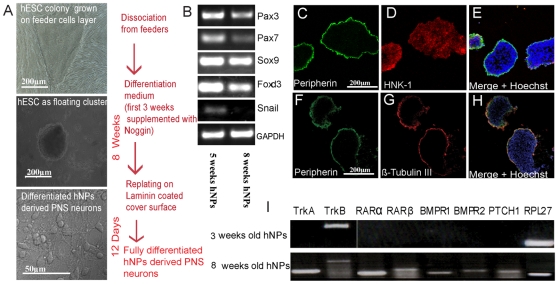
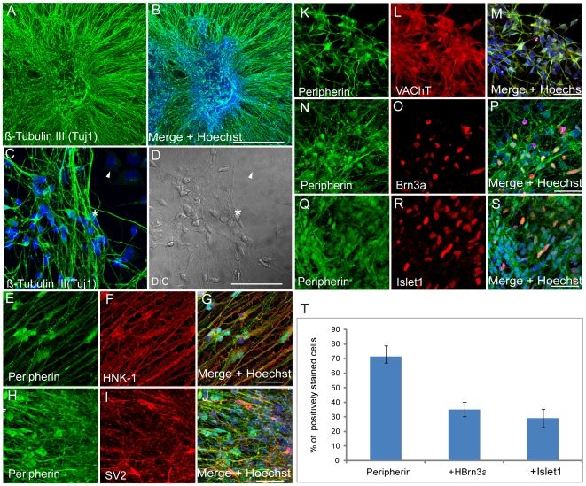
Background: The absence of a suitable cellular model is a major obstacle for the study of peripheral neuropathies. Human embryonic stem cells hold the potential to be differentiated into peripheral neurons which makes them a suitable candidate for this purpose. However, so far the potential of hESC to differentiate into derivatives of the peripheral nervous system (PNS) was not investigated enough and in particular, the few trials conducted resulted in low yields of PNS neurons. Here we describe a novel hESC differentiation method to produce enriched populations of PNS mature neurons. By plating 8 weeks hESC derived neural progenitors (hESC-NPs) on laminin for two weeks in a defined medium, we demonstrate that over 70% of the resulting neurons express PNS markers and 30% of these cells are sensory neurons.
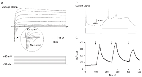
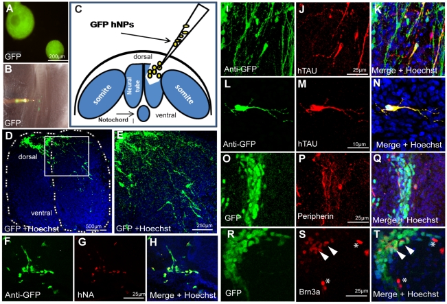
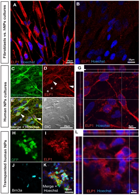
Methods/findings: Our method shows that the hNPs express neuronal crest lineage markers in a temporal manner, and by plating 8 weeks hESC-NPs into laminin coated dishes these hNPs were promoted to differentiate and give rise to homogeneous PNS neuronal populations, expressing several PNS lineage-specific markers. Importantly, these cultures produced functional neurons with electrophysiological activities typical of mature neurons. Moreover, supporting this physiological capacity implantation of 8 weeks old hESC-NPs into the neural tube of chick embryos also produced human neurons expressing specific PNS markers in vivo in just a few days. Having the enriched PNS differentiation system in hand, we show for the first time in human PNS neurons the expression of IKAP/hELP1 protein, where a splicing mutation on the gene encoding this protein causes the peripheral neuropathy Familial Dysautonomia.
Conclusions/significance: We conclude that this differentiation system to produce high numbers of human PNS neurons will be useful for studying PNS related neuropathies and for developing future drug screening applications for these diseases.
Conflict of interest statement
Figures
Similar articles
-
Familial Dysautonomia (FD) Human Embryonic Stem Cell Derived PNS Neurons Reveal that Synaptic Vesicular and Neuronal Transport Genes Are Directly or Indirectly Affected by IKBKAP Downregulation.PLoS One. 2015 Oct 5;10(10):e0138807. doi: 10.1371/journal.pone.0138807. eCollection 2015. PLoS One. 2015. PMID: 26437462 Free PMC article.
-
PA6-induced human embryonic stem cell-derived neurospheres: a new source of human peripheral sensory neurons and neural crest cells.Brain Res. 2008 Sep 16;1230:50-60. doi: 10.1016/j.brainres.2008.07.029. Epub 2008 Jul 16. Brain Res. 2008. PMID: 18671952
-
Involvement of IKAP in peripheral target innervation and in specific JNK and NGF signaling in developing PNS neurons.PLoS One. 2014 Nov 19;9(11):e113428. doi: 10.1371/journal.pone.0113428. eCollection 2014. PLoS One. 2014. PMID: 25409162 Free PMC article.
-
[Boundary cap cells--a nest of neural stem cells in the peripheral nervous system].Bull Acad Natl Med. 2007 Oct;191(7):1383-92; discussion 1392-4. Bull Acad Natl Med. 2007. PMID: 18447060 Review. French.
-
Lateral neural borders as precursors of peripheral nervous systems: A comparative view across bilaterians.Dev Growth Differ. 2019 Jan;61(1):58-72. doi: 10.1111/dgd.12585. Epub 2018 Dec 21. Dev Growth Differ. 2019. PMID: 30575021 Review.
Cited by
-
Altered Secretome and ROS Production in Olfactory Mucosa Stem Cells Derived from Friedreich's Ataxia Patients.Int J Mol Sci. 2020 Sep 11;21(18):6662. doi: 10.3390/ijms21186662. Int J Mol Sci. 2020. PMID: 32933002 Free PMC article.
-
Olfactory stem cells, a new cellular model for studying molecular mechanisms underlying familial dysautonomia.PLoS One. 2010 Dec 20;5(12):e15590. doi: 10.1371/journal.pone.0015590. PLoS One. 2010. PMID: 21187979 Free PMC article.
-
Induced pluripotent stem cells for disease modeling, cell therapy and drug discovery in genetic autonomic disorders: a review.Clin Auton Res. 2019 Aug;29(4):367-384. doi: 10.1007/s10286-018-00587-4. Epub 2019 Jan 10. Clin Auton Res. 2019. PMID: 30631982 Review.
-
A Novel In Vitro Assay Using Human iPSC-Derived Sensory Neurons to Evaluate the Effects of External Chemicals on Neuronal Morphology: Possible Implications in the Prediction of Abnormal Skin Sensation.Int J Mol Sci. 2021 Sep 29;22(19):10525. doi: 10.3390/ijms221910525. Int J Mol Sci. 2021. PMID: 34638865 Free PMC article.
-
Selective Induction of Human Autonomic Neurons Enables Precise Control of Cardiomyocyte Beating.Sci Rep. 2020 Jun 11;10(1):9464. doi: 10.1038/s41598-020-66303-3. Sci Rep. 2020. PMID: 32528170 Free PMC article.
References
-
- Zeng X, Rao MS. Human Embryonic Stem Cells: long term stability, absence of senescence and a potential cell source for neural replacement. Neuroscience. 2007;145:1348–1358. - PubMed
-
- Reubinoff B, Pera M, Fong C, Trounson A, Bongso A. Embryonic stem cell lines from human blastocysts: somatic differentiation in vitro. Nat Biotechnol. 2000;18:399–404. - PubMed
-
- Reubinoff BE IP, Turetsky T, Pera MF, Reinhartz E, Itzik A, et al. Neural progenitors from human embryonic stem cells. Nature Biotechnology. 2001;19:1134–1140. - PubMed
-
- Itsykson P, Ilouz N, Turetsky T, Goldstein RS, Pera MF, et al. Derivation of neural precursors from human embryonic stem cells in the presence of noggin. Mol Cell Neurosci. 2005;30:24–36. - PubMed
-
- Zhang S, Wernig M, Duncan I, Brüstle O, Thomson J. In vitro differentiation of transplantable neural precursors from human embryonic stem cells. Nat Biotechnol. 2001;19:1129–1133. - PubMed
Publication types
MeSH terms
Substances
LinkOut - more resources
Full Text Sources
Other Literature Sources
Medical
