

Therapeutic efficacy of human hepatocyte transplantation in a SCID/uPA mouse model with inducible liver disease
- PMID: 20174638
- PMCID: PMC2823785
- DOI: 10.1371/journal.pone.0009209
Therapeutic efficacy of human hepatocyte transplantation in a SCID/uPA mouse model with inducible liver disease
Abstract
Background: Severe Combined Immune Deficient (SCID)/Urokinase-type Plasminogen Activator (uPA) mice undergo liver failure and are useful hosts for the propagation of transplanted human hepatocytes (HH) which must compete with recipient-derived hepatocytes for replacement of the diseased liver parenchyma. While partial replacement by HH has proven useful for studies with Hepatitis C virus, complete replacement of SCID/uPA mouse liver by HH has never been achieved and limits the broader application of these mice for other areas of biomedical research. The herpes simplex virus type-1 thymidine kinase (HSVtk)/ganciclovir (GCV) system is a powerful tool for cell-specific ablation in transgenic animals. The aim of this study was to selectively eliminate murine-derived parenchymal liver cells from humanized SCID/uPA mouse liver in order to achieve mice with completely humanized liver parenchyma. Thus, we reproduced the HSVtk (vTK)/GCV system of hepatic failure in SCID/uPA mice.
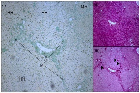
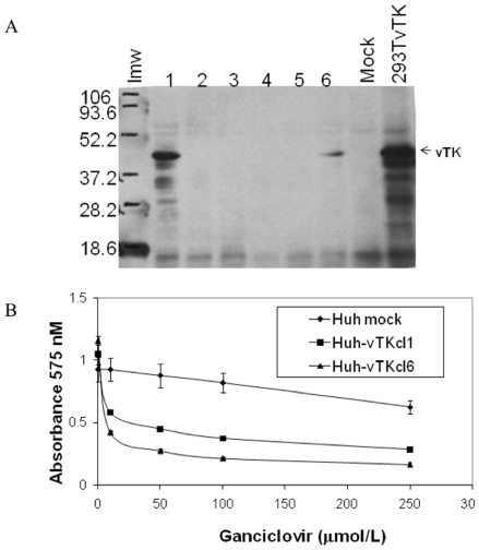
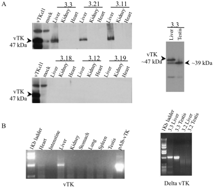
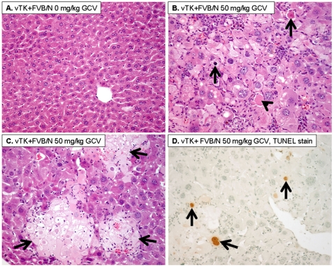
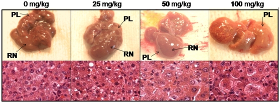
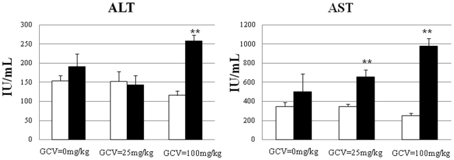
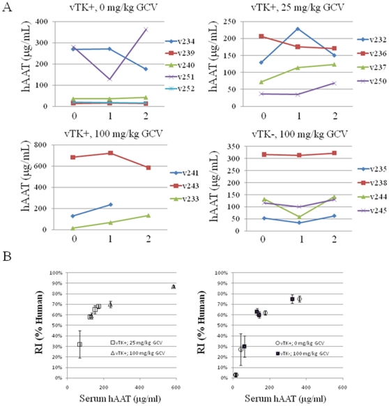
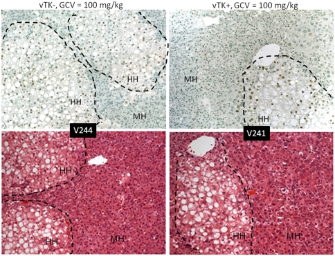
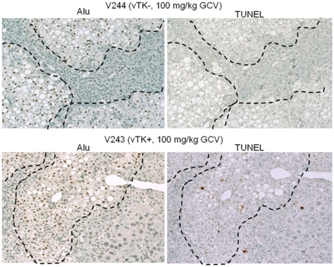
Methodology/principal findings: In vitro experiments demonstrated efficient killing of vTK expressing hepatoma cells after GCV treatment. For in vivo experiments, expression of vTK was targeted to the livers of FVB/N and SCID/uPA mice. Hepatic sensitivity to GCV was first established in FVB/N mice since these mice do not undergo liver failure inherent to SCID/uPA mice. Hepatic vTK expression was found to be an integral component of GCV-induced pathologic and biochemical alterations and caused death due to liver dysfunction in vTK transgenic FVB/N and non-transplanted SCID/uPA mice. In SCID/uPA mice with humanized liver, vTK/GCV caused death despite extensive replacement of the mouse liver parenchyma with HH (ranging from 32-87%). Surprisingly, vTK/GCV-dependent apoptosis and mitochondrial aberrations were also localized to bystander vTK-negative HH.
Conclusions/significance: Extensive replacement of mouse liver parenchyma by HH does not provide a secure therapeutic advantage against vTK/GCV-induced cytotoxicity targeted to residual mouse hepatocytes. Functional support by engrafted HH may be secured by strategies aimed at limiting this bystander effect.
Conflict of interest statement
Figures

Similar articles
-
A novel TK-NOG based humanized mouse model for the study of HBV and HCV infections.Biochem Biophys Res Commun. 2013 Nov 8;441(1):230-5. doi: 10.1016/j.bbrc.2013.10.040. Epub 2013 Oct 16. Biochem Biophys Res Commun. 2013. PMID: 24140055
-
Propagation of Human Hepatocytes in uPA/SCID Mice: Producing Chimeric Mice with Humanized Liver.Methods Mol Biol. 2017;1506:91-100. doi: 10.1007/978-1-4939-6506-9_6. Methods Mol Biol. 2017. PMID: 27830547
-
A mouse model of inducible liver injury caused by tet-on regulated urokinase for studies of hepatocyte transplantation.Am J Pathol. 2009 Nov;175(5):1975-83. doi: 10.2353/ajpath.2009.090349. Epub 2009 Oct 1. Am J Pathol. 2009. PMID: 19808649 Free PMC article.
-
The human liver-uPA-SCID mouse: a model for the evaluation of antiviral compounds against HBV and HCV.Antiviral Res. 2008 Dec;80(3):231-8. doi: 10.1016/j.antiviral.2008.07.006. Epub 2008 Aug 14. Antiviral Res. 2008. PMID: 18706933 Review.
-
Mice with human livers.Gastroenterology. 2013 Dec;145(6):1209-14. doi: 10.1053/j.gastro.2013.09.009. Epub 2013 Sep 13. Gastroenterology. 2013. PMID: 24042096 Review.
Cited by
-
Cell therapy for liver disorders: past, present and future.Nat Rev Gastroenterol Hepatol. 2025 May;22(5):329-342. doi: 10.1038/s41575-025-01050-2. Epub 2025 Mar 18. Nat Rev Gastroenterol Hepatol. 2025. PMID: 40102584 Review.
-
Usage of adenovirus expressing thymidine kinase mediated hepatocellular damage for enabling mouse liver repopulation with allogenic or xenogenic hepatocytes.PLoS One. 2013 Sep 24;8(9):e74948. doi: 10.1371/journal.pone.0074948. eCollection 2013. PLoS One. 2013. PMID: 24086405 Free PMC article.
-
Murine Models of Hepatitis A Virus Infection.Cold Spring Harb Perspect Med. 2019 Jan 2;9(1):a031674. doi: 10.1101/cshperspect.a031674. Cold Spring Harb Perspect Med. 2019. PMID: 29661811 Free PMC article. Review.
-
Oxidative Stress Attenuates Lipid Synthesis and Increases Mitochondrial Fatty Acid Oxidation in Hepatoma Cells Infected with Hepatitis C Virus.J Biol Chem. 2016 Jan 22;291(4):1974-1990. doi: 10.1074/jbc.M115.674861. Epub 2015 Dec 1. J Biol Chem. 2016. PMID: 26627833 Free PMC article.
-
The nude mouse as model for liver deficiency study and treatment and xenotransplantation.Int J Hepatol. 2012;2012:140147. doi: 10.1155/2012/140147. Epub 2012 Oct 31. Int J Hepatol. 2012. PMID: 23193481 Free PMC article.
References
-
- Rodrigues AD. Comparison of levels of aldehyde oxidase with cytochrome P450 activities in human liver in vitro. Biochem Pharmacol. 1994;48:197–200. - PubMed
-
- Rodrigues AD, Kukulka MJ, Surber BW, Thomas SB, Uchic JT, et al. Measurement of liver microsomal cytochrome p450 (CYP2D6) activity using [O-methyl-14C]dextromethorphan. Anal Biochem. 1994;219:309–320. - PubMed
-
- Rodrigues AD, Wong SL. Application of human liver microsomes in metabolism-based drug-drug interactions: in vitro-in vivo correlations and the Abbott Laboratories experience. Adv Pharmacol. 1997;43:65–101. - PubMed
-
- Barry M, Feely J. Enzyme induction and inhibition. Pharmacol Ther. 1990;48:71–94. - PubMed
-
- Park BK, Breckenridge AM. Clinical implications of enzyme induction and enzyme inhibition. Clin Pharmacokinet. 1981;6:1–24. - PubMed
Publication types
MeSH terms
Substances
Grants and funding
LinkOut - more resources
Full Text Sources
Medical
Miscellaneous
