. 2010 Feb 17;5(2):e9261.
doi: 10.1371/journal.pone.0009261.

Developmental sex differences in nicotinic currents of prefrontal layer VI neurons in mice and rats

Affiliations

Developmental sex differences in nicotinic currents of prefrontal layer VI neurons in mice and rats

Nyresa C Alves et al. PLoS One. .

Abstract

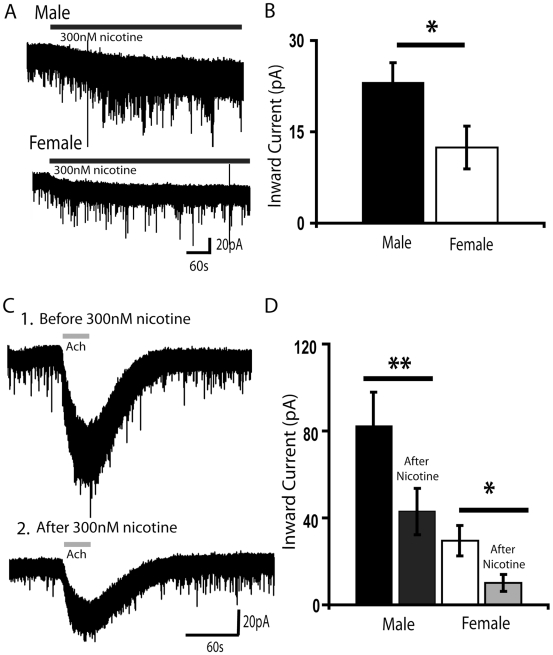
Background: There is a large sex difference in the prevalence of attention deficit disorder; yet, relatively little is known about sex differences in the development of prefrontal attention circuitry. In male rats, nicotinic acetylcholine receptors excite corticothalamic neurons in layer VI, which are thought to play an important role in attention by gating the sensitivity of thalamic neurons to incoming stimuli. These nicotinic currents in male rats are significantly larger during the first postnatal month when prefrontal circuitry is maturing. The present study was undertaken to investigate whether there are sex differences in the nicotinic currents in prefrontal layer VI neurons during development.

Methodology/principal findings: Using whole cell recording in prefrontal brain slice, we examined the inward currents elicited by nicotinic stimulation in male and female rats and two strains of mice. We found a prominent sex difference in the currents during the first postnatal month when males had significantly greater nicotinic currents in layer VI neurons compared to females. These differences were apparent with three agonists: acetylcholine, carbachol, and nicotine. Furthermore, the developmental sex difference in nicotinic currents occurred despite male and female rodents displaying a similar pattern and proportion of layer VI neurons possessing a key nicotinic receptor subunit.

Conclusions/significance: This is the first illustration at a cellular level that prefrontal attention circuitry is differently affected by nicotinic receptor stimulation in males and females during development. This transient sex difference may help to define the cellular and circuit mechanisms that underlie vulnerability to attention deficit disorder.


Conflict of interest statement

Competing Interests: The authors have declared that no competing interests exist.

Figures

Figure 1
Figure 1. Developmental sex difference in nicotinic currents in layer VI neurons of prefrontal cortex.
(A) Examples of voltage clamp traces from a P19 male and a P27 female showing nicotinic inward currents during bath application of acetylcholine (1 mM, 10 s). Line denotes acetylcholine application. Both males and females have reproducible, non-desensitizing currents elicited by bath-applied acetylcholine, when given five-minute washout duration. (B) Bar chart summarizing the mean amplitude of the peak inward current elicited by acetylcholine in FVB male (left panel) and female (right panel) mice in layer VI across postnatal weeks two to five. In males, there is a significant developmental effect where the mean nicotinic current during postnatal weeks three and four are significantly higher than the mean inward current during postnatal weeks two and five (* P<0.05). In females, there is also a significant developmental effect where the mean nicotinic current during postnatal week three is significantly higher than the mean inward current during postnatal weeks two and five (* P<0.05). (C) Bar graph displays the sex difference in the average inward current elicited by nicotinic receptor stimulation by acetylcholine (1 mM, 30 s). Males (black bars) have significantly greater currents than females (open bars) during postnatal weeks three and four (** P<0.01). All recordings are performed in the presence of atropine (200 nM) to block muscarinic receptors and MLA (10 nM) to block α7 nicotinic receptors.
Figure 2
Figure 2. Developmental sex difference in nicotinic currents is not explained by different levels of acetylcholinesterase activity.
(A) Voltage clamp traces showing inward currents during bath application of nicotinic acetylcholine receptor agonists (1) acetylcholine (1 mM, 30 s) and (2) carbachol (1 mM, 30 s), an acetylcholine analogue that is not broken down by endogenous acetylcholinesterase, in the same layer VI neuron from a P17 male FVB mouse. (B) Voltage clamp traces from the same agonist applications in a layer VI neuron from a P17 female FVB mouse. In both, males and females, the inward current persists longer after carbachol compared to acetylcholine, since the acetylcholinesterase in the brain slice metabolizes applied acetylcholine allowing the cell to return to baseline faster. (C) Bar chart summarizing the mean current amplitude elicited by 30 s application of 1 mM acetylcholine or carbachol (**P<0.01). The sex difference persists when the inward currents are elicited with 1 mM carbachol, suggesting that acetylcholinesterase levels do not account for the sex difference in nicotinic currents.
Figure 3
Figure 3. Developmental sex difference in current elicited by nicotine, but not its desensitization of acetylcholine currents.
(A) Exemplary voltage-clamp traces showing a small, persistent inward current elicited by nicotine (300 nM, 10 min) in a layer VI neuron from a P21 male (top) and a P19 female (bottom). This concentration of nicotine is consistent with the peak blood level of nicotine seen in smokers and is relevant to developmental nicotine exposure . (B) Bar graph to the right showing the mean inward current elicited by 300 nM nicotine in typical male and female layer VI neurons. Nicotine elicited greater inward currents in male neurons than females (P<0.05). (C 1) A voltage-clamp trace from a P21 male shows a robust inward current with acetylcholine (1 mM, 30 s) before application of nicotine. (C 2) A voltage-clamp trace from the same neuron taken five minutes after the end of a ten minute application of nicotine (300 nM) shows that the inward current elicited by acetylcholine (1 mM, 30 s) is significantly decreased. (D) Bar chart showing the highly significant suppression of the inward current elicited by acetylcholine (1 mM, 30 s) in males and females after the above nicotine exposure. The latter inward currents elicited by acetylcholine were examined five minutes after nicotine application when its inward current had returned to baseline (*P<0.05, ** P<0.01).
Figure 4
Figure 4. Developmental nicotinic currents and nicotinic α4YFP-positive neurons in male and female knock-in mice.
(A) Bar graph showing larger inward currents in male α4YFP knock-in mice compared to age-matched female α4YFP knock-in mice during postnatal week three (**P<0.01). (B) Low-magnification image of P15 male and female prefrontal cortex slices with the YFP signal on the α4YFP subunits amplified using a 3-step immunohistochemistry protocol described in the Methods section. Both sexes show a distinct neuronal band of staining in layer VI of the medial prefrontal cortex (bright red cells), showing the presence of α4* nicotinic receptors. Scale bar: 200 µm. (C) High-magnification of (1) YFP immunostained neurons, (2) DAPI stained cells, and (3) merged images within layer VI of male (top images) and female (bottom images) prefrontal cortex. The criteria for identifying DAPI-positive neurons are described in the Methods section. The proportion of neurons expressing α4YFP was not significantly different between males and females. Scale bar: 20 µm.

References

    1. Brown RT, Freeman WS, Perrin JM, Stein MT, Amler RW, et al. Prevalence and assessment of attention-deficit/hyperactivity disorder in primary care settings. Pediatrics. 2001;107:E43. - PubMed
    1. Cuffe SP, Moore CG, McKeown RE. Prevalence and correlates of ADHD symptoms in the national health interview survey. Journal of attention disorders. 2005;9:392–401. - PubMed
    1. Smalley SL, McGough JJ, Moilanen IK, Loo SK, Taanila A, et al. Prevalence and psychiatric comorbidity of attention-deficit/hyperactivity disorder in an adolescent Finnish population. Journal of the American Academy of Child and Adolescent Psychiatry. 2007;46:1575–1583. - PubMed
    1. Shaw P, Eckstrand K, Sharp W, Blumenthal J, Lerch JP, et al. Attention-deficit/hyperactivity disorder is characterized by a delay in cortical maturation. Proc Natl Acad Sci USA. 2007;104:19649–19654. - PMC - PubMed
    1. Sullivan RM, Brake WG. What the rodent prefrontal cortex can teach us about attention-deficit/hyperactivity disorder: the critical role of early developmental events on prefrontal function. Behav Brain Res. 2003;146:43–55. - PubMed

Publication types

MeSH terms