

Engineered heart tissue: a novel tool to study the ischemic changes of the heart in vitro
- PMID: 20174664
- PMCID: PMC2822866
- DOI: 10.1371/journal.pone.0009275
Engineered heart tissue: a novel tool to study the ischemic changes of the heart in vitro
Abstract
Background: Understanding the basic mechanisms and prevention of any disease pattern lies mainly on development of a successful experimental model. Recently, engineered heart tissue (EHT) has been demonstrated to be a useful tool in experimental transplantation. Here, we demonstrate a novel function for the spontaneously contracting EHT as an experimental model in studying the acute ischemia-induced changes in vitro.
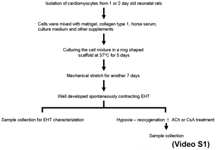
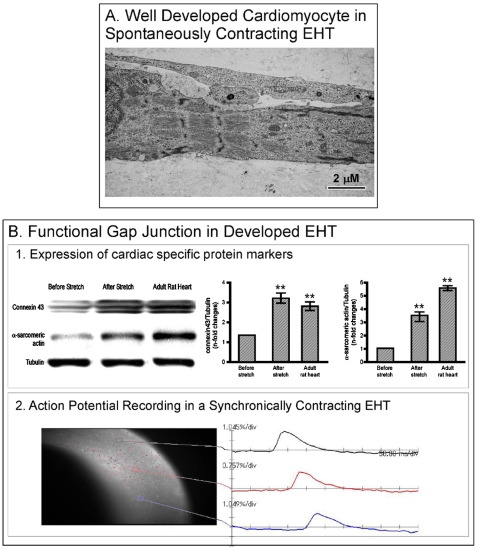
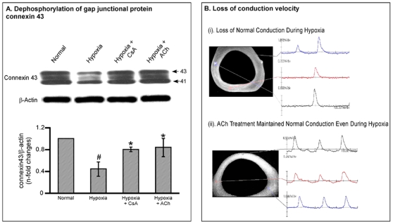
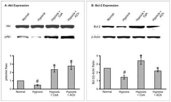
Methodology/principal findings: EHT was constructed by mixing cardiomyocytes isolated from the neonatal rats and cultured in a ring-shaped scaffold for five days. This was followed by mechanical stretching of the EHT for another one week under incubation. Fully developed EHT was subjected to hypoxia with 1% O(2) for 6 hours after treating them with cell protective agents such as cyclosporine A (CsA) and acetylcholine (ACh). During culture, EHT started to show spontaneous contractions that became more synchronous following mechanical stretching. This was confirmed by the increased expression of gap junctional protein connexin 43 and improved action potential recordings using an optical mapping system after mechanical stretching. When subjected to hypoxia, EHT demonstrated conduction defects, dephosphorylation of connexin-43, and down-regulation of cell survival proteins identical to the adult heart. These effects were inhibited by treating the EHT with cell protective agents.
Conclusions/significance: Under hypoxic conditions, the EHT responds similarly to the adult myocardium, thus making EHT a promising material for the study of cardiac functions in vitro.
Conflict of interest statement
Figures
Similar articles
-
Functional improvement and maturation of rat and human engineered heart tissue by chronic electrical stimulation.J Mol Cell Cardiol. 2014 Sep;74:151-61. doi: 10.1016/j.yjmcc.2014.05.009. Epub 2014 May 19. J Mol Cell Cardiol. 2014. PMID: 24852842
-
Terminal differentiation, advanced organotypic maturation, and modeling of hypertrophic growth in engineered heart tissue.Circ Res. 2011 Oct 28;109(10):1105-14. doi: 10.1161/CIRCRESAHA.111.251843. Epub 2011 Sep 15. Circ Res. 2011. PMID: 21921264
-
Transplantation of engineered heart tissue as a biological cardiac assist device for treatment of dilated cardiomyopathy.Eur J Heart Fail. 2013 Jan;15(1):23-35. doi: 10.1093/eurjhf/hfs200. Eur J Heart Fail. 2013. PMID: 23243122
-
Engineered heart tissue for regeneration of diseased hearts.Biomaterials. 2004 Apr;25(9):1639-47. doi: 10.1016/s0142-9612(03)00521-0. Biomaterials. 2004. PMID: 14697865 Review.
-
Cardiac tissue engineering.Transpl Immunol. 2002 May;9(2-4):315-21. doi: 10.1016/s0966-3274(02)00011-4. Transpl Immunol. 2002. PMID: 12180846 Review.
Cited by
-
Protection by the NO-Donor SNAP and BNP against Hypoxia/Reoxygenation in Rat Engineered Heart Tissue.PLoS One. 2015 Jul 6;10(7):e0132186. doi: 10.1371/journal.pone.0132186. eCollection 2015. PLoS One. 2015. PMID: 26147889 Free PMC article.
-
Multicellular 3D models to study myocardial ischemia-reperfusion injury.Front Cell Dev Biol. 2024 Nov 15;12:1494911. doi: 10.3389/fcell.2024.1494911. eCollection 2024. Front Cell Dev Biol. 2024. PMID: 39620142 Free PMC article. Review.
-
Disease-inspired tissue engineering: Investigation of cardiovascular pathologies.ACS Biomater Sci Eng. 2020 May 11;6(5):2518-2532. doi: 10.1021/acsbiomaterials.9b01067. Epub 2019 Oct 29. ACS Biomater Sci Eng. 2020. PMID: 32974421 Free PMC article. Review.
-
Cardiovascular Organ-on-a-Chip Platforms for Drug Discovery and Development.Appl In Vitro Toxicol. 2016 Jun 1;2(2):82-96. doi: 10.1089/aivt.2016.0002. Appl In Vitro Toxicol. 2016. PMID: 28971113 Free PMC article. Review.
-
A combined synthetic-fibrin scaffold supports growth and cardiomyogenic commitment of human placental derived stem cells.PLoS One. 2012;7(4):e34284. doi: 10.1371/journal.pone.0034284. Epub 2012 Apr 3. PLoS One. 2012. PMID: 22509287 Free PMC article.
References
-
- Zimmermann WH, Fink C, Kralisch D, Remmers U, Weil J, et al. Three-dimensional engineered heart tissue from neonatal rat cardiac myocytes. Biotechnol Bioeng. 2000;68:106–114. - PubMed
-
- Zimmermann WH, Melnychenko I, Wasmeier G, Didie M, Naito H, et al. Engineered heart tissue grafts improve systolic and diastolic function in infarcted rat hearts. Nat Med. 2006;12:452–458. - PubMed
-
- Naito H, Melnychenko I, Didie M, Schneiderbanger K, Schubert P, et al. Optimizing engineered heart tissue for therapeutic applications as surrogate heart muscle. Circulation. 2006;114:I72–78. - PubMed
-
- Zimmermann WH, Schneiderbanger K, Schubert P, Didie M, Munzel F, et al. Tissue engineering of a differentiated cardiac muscle construct. Circ Res. 2002;90:223–230. - PubMed
Publication types
MeSH terms
Substances
LinkOut - more resources
Full Text Sources
Other Literature Sources
Miscellaneous
