Review
. 2010 Mar;135(3):441-51.
doi: 10.1039/b907735a. Epub 2009 Dec 22.

Single molecule sensing by nanopores and nanopore devices

Affiliations
Review

Single molecule sensing by nanopores and nanopore devices

Li-Qun Gu et al. Analyst. 2010 Mar.

Abstract

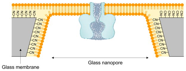
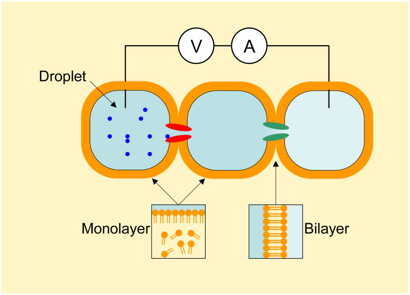
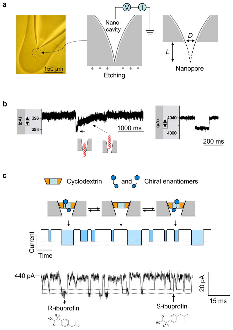
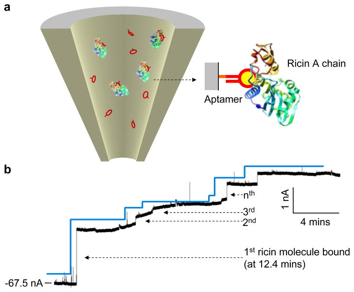
Molecular-scale pore structures, called nanopores, can be assembled by protein ion channels through genetic engineering or be artificially fabricated on solid substrates using fashion nanotechnology. When target molecules interact with the functionalized lumen of a nanopore, they characteristically block the ion pathway. The resulting conductance changes allow for identification of single molecules and quantification of target species in the mixture. In this review, we first overview nanopore-based sensory techniques that have been created for the detection of myriad biomedical targets, from metal ions, drug compounds, and cellular second messengers to proteins and DNA. Then we introduce our recent discoveries in nanopore single molecule detection: (1) using the protein nanopore to study folding/unfolding of the G-quadruplex aptamer; (2) creating a portable and durable biochip that is integrated with a single-protein pore sensor (this chip is compared with recently developed protein pore sensors based on stabilized bilayers on glass nanopore membranes and droplet interface bilayer); and (3) creating a glass nanopore-terminated probe for single-molecule DNA detection, chiral enantiomer discrimination, and identification of the bioterrorist agent ricin with an aptamer-encoded nanopore.


Figures

Figure 1
Figure 1
Single-molecule detection in a protein pore. (a) Competitive and reversible binding of different analytes (represented by green and red balls) to a receptor engineered in the protein pore. (b) Stochastic current blocks function as “signatures” of single bound analytes, which, based on block amplitude and duration, allow for the identification of the unknown analytes. In the diagram, the red analyte blocks more current with a shorter duration than the green one. Analytes can also be quantified by their block occurrence.
Figure 2
Figure 2
Molecular graph of the heptameric α-hemolysin pore in the lipid bilayer and diagrams for various single molecule detections using α-hemolysin. The dimensions of the various regions in the lumen of the pore are provided.
Figure 3
Figure 3
Examining the folding and unfolding of a single G-quadruplex aptamer in the nanocavity of the αHL pore. (a) The sequence and structure of the G-quadruplex formed by the thrombin-binding aptamer (TBA) (left) and the two G-tetrad planes in the TBA G-quadruplex (right). The top tetrad is formed by guanines at the positions 1, 6, 10 and 15, and the bottom is formed by guanine 2, 5, 11 and 14. A cation in between is coordinated with eight carbonyls. The average cation-carbonyl distance is 2.86 Å. (b) Current block signal. (c) The long-lived block for capturing a single G-quadruplex in the nanocavity enclosed by the αHL pore. (d) The spike at the long block terminal produced by translocation of an unfolded G-quadruplex in the nanocavity. (e) Short-lived blocks formed by translocation of the linear form of TBA. (g) Characteristic blocks produced by tag-TBA (top) and the model showing the molecular location and position in the cavity (bottom). (h) Another type of block by tag-TBA (top) and the corresponding model showing the change in position of the molecule (bottom). This figure has been modified from references,.
Figure 4
Figure 4
Fabrication, prototype and use of the modular ion channel chip. (a) Assembly of the chip. The analyte can be added from the sample cell on the back of either compartment and delivered to the sensor element through agarose layered between the sample cell and the membrane. (b) A device prototype. (c) Signature blocks by the second messenger IP3 (500 nM) on the chip in a simulated intracellular conditions: 150 mM KCl, 2 mM ATP, 2.3 mM MgCl2, 10 mM Tris, 0.3 mM ATP, pH 7.4. This figure has been modified from reference.
Figure 5
Figure 5
Incorporation of the protein pore in the lipid bilayer suspended over a glass nanopore (GNP) membrane. The GNP is a single conical-shaped nanopore embedded in a 50 μm glass membrane. The fabrication of GNP membrane was described in the reference. The interior and exterior surfaces of the GNP are coated with 3-cyanopropyldimethylchlorosilane to form a hydrophobic surface, which allows the formation of lipid monolayer on it. Lipid monolayers on the interior and exterior surfaces of the GNP will merge to form an exceptionally stable bilayer across the GNP.
Figure 6
Figure 6
Droplet interface bilayer (DIB) and DIB network. The DIB is formed on the interface between two aqueous droplets in the lipid solution. Firstly, a liquid droplet is created by dipping a drop of aqueous solution into the lipid solution, with a lipid monolayer formed on the droplet-oil interface. Then two droplets are manipulated to contact each other and their surface lipid monolayers will hybridize into a bilayer on the interface, DIB. Various channel proteins in the droplet solution can be incorporated into the DIB, and recorded using two Ag/AgCl electrodes, each of which is inserted into a droplet. A very useful property of DIB is the ability to form network, in which each DIB contains a responsive protein channel. The DIB network acts as a molecular device to simulate functions of semiconductor circuits, such as half-wave rectification and full-wave rectification. The droplets can also form arrays for parallel screening ion channel blocks.
Figure 7
Figure 7
Glass nanopore-terminated probe and single molecule manipulation. (a) The glass nanopore is fabricated by sealing the micropipette terminal to enclose a nanocavity, followed by external etching the glass terminal with electrical monitoring to perforate the nanocavity with controllable pore size. (b) Current blocks showing translocation of 1 kbp dsDNA through a 2 nm nanopore (left) and a 7 nm pore (right) in 1 M NaCl and recorded +100 mV. When the pore size is comparable to dsDNA, the DNA translocation speed is slowed down and translocation steps can be revealed from the block type (left), which is different from that for DNA translocation in a wider pore (right). (c) Single molecule discrimination of chiral enantiomers by the cyclodextrin (1.5 nm) trapped in the 1 nm nanopore. The interaction of chiral compounds with cyclodextrin can be separated from their block durations and current amplitudes. The current trace showed the binding of individual enantiomers of ibuprofen to the trapped β-cyclodextrin in the nanopore. The solution contained the mixture of 100 μM R-(−)-ibuprofen and 100 μM S-(+)-ibuprofen. The current block levels by R- and S-ibuprofen were marked with dash lines.
Figure 8
Figure 8
Single-molecule detection of bioterrorist agent ricin with an aptamer-encoded nanopore. The ricin A chain-targeted aptamer was immobilized on the inner surface of the glass nanopore. The current trace showed a 56 nm aptamer-encoded nanopore in the presence of 100 nM ricin A-chain protein in the external solution (−100 mV). The stepwise current blocks are attributed to the sequential binding of single ricin molecules to the immobilized aptamers in the nanopore.

Similar articles

Cited by

References

    1. B. Hille, 2001, 3rd,

    1. Bayley H, Cremer PS. Nature. 2001;413:226–230. - PubMed
    1. Song LZ, Hobaugh MR, Shustak C, Cheley S, Bayley H, Gouaux JE. Science. 1996;274:1859–1866. - PubMed
    1. Bayley H, Cremer PS. Nature. 2001;413:226–230. - PubMed
    1. Bayley H, Jayasinghe L. Molecular Membrane Biology. 2004;21:209–220. - PubMed

Publication types