

Protein scaffold-based molecular probes for cancer molecular imaging
- PMID: 20174842
- PMCID: PMC2914822
- DOI: 10.1007/s00726-010-0503-9
Protein scaffold-based molecular probes for cancer molecular imaging
Abstract
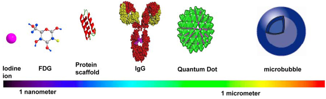
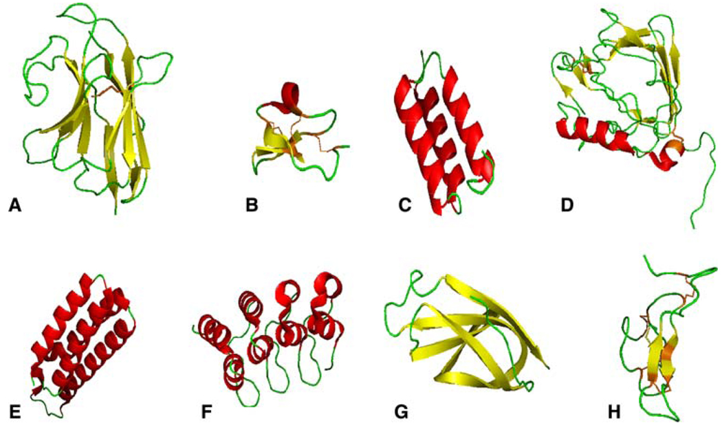
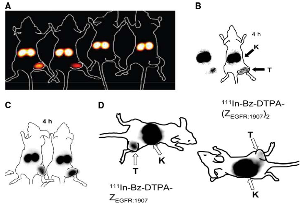
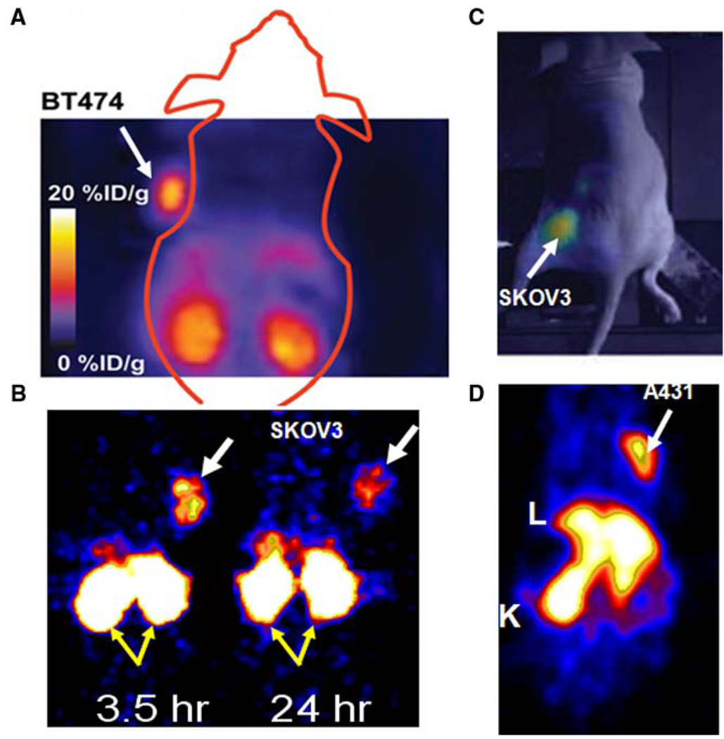
Protein scaffold molecules are powerful reagents for targeting various cell signal receptors, enzymes, cytokines and other cancer-related molecules. They belong to the peptide and small protein platform with distinct properties. For the purpose of development of new generation molecular probes, various protein scaffold molecules have been labeled with imaging moieties and evaluated both in vitro and in vivo. Among the evaluated probes Affibody molecules and analogs, cystine knot peptides, and nanobodies have shown especially good characteristics as protein scaffold platforms for development of in vivo molecular probes. Quantitative data obtained from positron emission tomography, single photon emission computed tomography/CT, and optical imaging together with biodistribution studies have shown high tumor uptakes and high tumor-to-blood ratios for these probes. High tumor contrast imaging has been obtained within 1 h after injection. The success of those molecular probes demonstrates the adequacy of protein scaffold strategy as a general approach in molecular probe development.
Figures
Similar articles
-
Protein-based tumor molecular imaging probes.Amino Acids. 2011 Nov;41(5):1013-36. doi: 10.1007/s00726-010-0545-z. Epub 2010 Mar 17. Amino Acids. 2011. PMID: 20232092 Free PMC article. Review.
-
111In/68Ga-Labeled anti-epidermal growth factor receptor, native chemical ligation cyclized Affibody ZHER2:342min.2013 Apr 3 [updated 2013 May 23]. In: Molecular Imaging and Contrast Agent Database (MICAD) [Internet]. Bethesda (MD): National Center for Biotechnology Information (US); 2004–2013. 2013 Apr 3 [updated 2013 May 23]. In: Molecular Imaging and Contrast Agent Database (MICAD) [Internet]. Bethesda (MD): National Center for Biotechnology Information (US); 2004–2013. PMID: 23700641 Free Books & Documents. Review.
-
Evaluation of a (64)Cu-labeled cystine-knot peptide based on agouti-related protein for PET of tumors expressing alphavbeta3 integrin.J Nucl Med. 2010 Feb;51(2):251-258. doi: 10.2967/jnumed.109.069831. J Nucl Med. 2010. PMID: 20124048 Free PMC article.
-
PET Imaging of HER2-Positive Tumors with Cu-64-Labeled Affibody Molecules.Mol Imaging Biol. 2019 Oct;21(5):907-916. doi: 10.1007/s11307-018-01310-5. Mol Imaging Biol. 2019. PMID: 30617730
-
Developing Targeted Hybrid Imaging Probes by Chelator Scaffolding.Bioconjug Chem. 2017 Jun 21;28(6):1722-1733. doi: 10.1021/acs.bioconjchem.7b00182. Epub 2017 May 10. Bioconjug Chem. 2017. PMID: 28462989 Free PMC article.
Cited by
-
Protein and peptide probes for molecular imaging.Amino Acids. 2011 Nov;41(5):1009-12. doi: 10.1007/s00726-011-0945-8. Amino Acids. 2011. PMID: 21643775 Free PMC article. No abstract available.
-
New strategy for monitoring targeted therapy: molecular imaging.Int J Nanomedicine. 2013;8:3703-13. doi: 10.2147/IJN.S51264. Epub 2013 Sep 30. Int J Nanomedicine. 2013. PMID: 24124361 Free PMC article. Review.
-
Axl and EGFR Dual-Specific Binding Affibody for Targeted Therapy in Nasopharyngeal Carcinoma.Cells. 2024 Nov 5;13(22):1823. doi: 10.3390/cells13221823. Cells. 2024. PMID: 39594573 Free PMC article.
-
The role of nuclear medicine in modern therapy of cancer.Tumour Biol. 2012 Jun;33(3):629-40. doi: 10.1007/s13277-012-0373-8. Epub 2012 Mar 24. Tumour Biol. 2012. PMID: 22446937 Review.
-
Preliminary evaluation of (177)Lu-labeled knottin peptides for integrin receptor-targeted radionuclide therapy.Eur J Nucl Med Mol Imaging. 2011 Apr;38(4):613-22. doi: 10.1007/s00259-010-1684-x. Epub 2010 Dec 10. Eur J Nucl Med Mol Imaging. 2011. PMID: 21153409
References
-
- Ahlgren S, Orlova A, Rosik D, Sandström M, Sjöberg A, Baastrup B, Widmark O, Fant G, Feldwisch J, Tolmachev V. Evaluation of maleimide derivative of DOTA for site-specific labeling of recombinant affibody molecules. Bioconjug Chem. 2008;19:235–243. - PubMed
-
- Ahlgren S, Wallberg H, Tran TA, Widström C, Hjertman M, Abrahmsén L, Berndorff D, Dinkelborg LM, Cyr JE, Feldwisch J, Orlova A, Tolmachev V. Targeting of HER2-expressing tumors with a site-specifically 99mTc-labeled recombinant affibody molecule, ZHER2:2395, with C-terminally engineered cysteine. J Nucl Med. 2009;50:781–789. - PubMed
-
- Amstutz P, Binz HK, Parizek P, Stumpp MT, Kohl A, Grutter MG, Forrer P, Plückthun A. Intracellular kinase inhibitors selected from combinatorial libraries of designed ankyrin repeat proteins. J Biol Chem. 2005;280:24715–24722. - PubMed
-
- Arbabi Ghahroudi M, Desmyter A, Wyns L, Hamers R, Muyldermans S. Selection and identification of single domain antibody fragments from camel heavy-chain antibodies. FEBS Lett. 1997;414:521–526. - PubMed
Publication types
MeSH terms
Substances
Grants and funding
LinkOut - more resources
Full Text Sources
Other Literature Sources
