

Radiofrequency ablation of osteoid osteoma in atypical locations: a case series
- PMID: 20174900
- PMCID: PMC2882005
- DOI: 10.1007/s11999-010-1265-0
Radiofrequency ablation of osteoid osteoma in atypical locations: a case series
Abstract
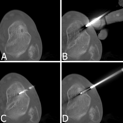
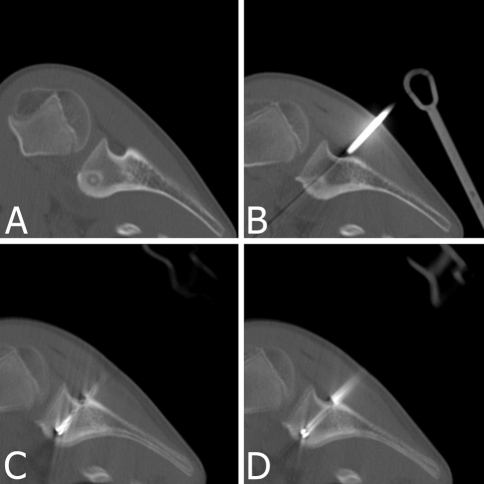
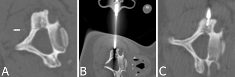
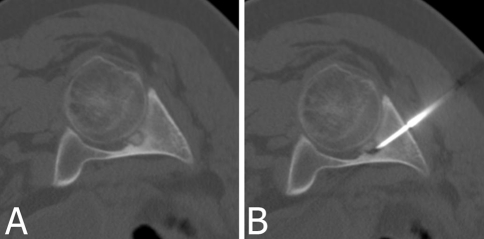
Background: Osteoid osteoma has a nidus surrounded by sclerotic bone with a size usually less than 20 mm. Its diagnosis is made on typical presentation of nocturnal pain and imaging findings. Excision of the niduses, which are often small and difficult to precisely identify, sometimes may result in resection of surrounding normal bone. Minimally invasive percutaneous treatments have been used to try to minimize resection of normal bone. Although minimally invasive radiofrequency ablation generally relieves pain, its ability to relieve pain is less well known in locations other than lower extremity long bones.
Questions/purposes: We determined the pain relief and complication rates after radiofrequency ablation of osteoid osteomas presenting in atypical locations and followed patients to assess possible recurrence or late complications.
Patients and methods: We retrospectively reviewed 21 patients with osteoid osteomas in unusual locations (eg, hip, radioulnar joint, and proximal phalanx) in whom we used radiofrequency ablation. Postoperative activities were not restricted for any of the patients. We assessed the time for patients to become symptom free, their activity status, and possible recurrence or complications. The minimum clinical followup was 12 months (mean, 27.8 months; range, 12-37 months).
Results: All patients became symptom free within 24 hours to 1 week. During followup, none of the patients experienced recurrence or any major complications.
Conclusions: Radiofrequency ablation for osteoid osteomas in unusual locations reliably relieves pain with few complications and recurrences at short-term followup.
Level of evidence: Level IV, case series. See Guidelines for Authors for a complete description of level of evidence.
Figures
Similar articles
-
Management of Osteoid Osteoma in Unusual Locations.Acta Chir Orthop Traumatol Cech. 2020;87(4):285-291. Acta Chir Orthop Traumatol Cech. 2020. PMID: 32940225 English.
-
Percutaneous Radiofrequency Ablation for the Treatment of Osteoid Osteoma in Children and Adults: A Comparative Analysis in 92 Patients.Cardiovasc Intervent Radiol. 2018 Sep;41(9):1384-1390. doi: 10.1007/s00270-018-1947-7. Epub 2018 Apr 12. Cardiovasc Intervent Radiol. 2018. PMID: 29651579
-
Case report: Osteoid osteoma of the acetabulum treated with arthroscopy-assisted radiofrequency ablation.Clin Orthop Relat Res. 2013 May;471(5):1727-32. doi: 10.1007/s11999-012-2772-y. Epub 2013 Jan 12. Clin Orthop Relat Res. 2013. PMID: 23315200 Free PMC article.
-
Comparison of arthroscopy versus percutaneous radiofrequency thermal ablation for the management of intra- and juxta-articular elbow osteoid osteoma: case series and a literature review.BMC Musculoskelet Disord. 2022 Mar 25;23(1):287. doi: 10.1186/s12891-022-05244-6. BMC Musculoskelet Disord. 2022. PMID: 35337326 Free PMC article. Review.
-
Percutaneous thermal ablation for treatment of osteoid osteoma: a systematic review and analysis.Skeletal Radiol. 2020 Sep;49(9):1403-1411. doi: 10.1007/s00256-020-03435-7. Epub 2020 Apr 8. Skeletal Radiol. 2020. PMID: 32270226
Cited by
-
Image-guided Percutaneous Radiofrequency Ablation for Osteoid Osteoma: Experience from a Developing Nation.Cureus. 2019 Sep 12;11(9):e5633. doi: 10.7759/cureus.5633. Cureus. 2019. PMID: 31700736 Free PMC article.
-
Computed tomography guided radio-frequency ablation of osteoid osteomas in atypical locations.Indian J Radiol Imaging. 2019 Jul-Sep;29(3):253-257. doi: 10.4103/ijri.IJRI_259_19. Epub 2019 Oct 30. Indian J Radiol Imaging. 2019. PMID: 31741592 Free PMC article.
-
Arthroscopic excision of acetabular osteoid osteoma in a 7-year-old patient.Knee Surg Sports Traumatol Arthrosc. 2015 Nov;23(11):3432-5. doi: 10.1007/s00167-014-2978-5. Epub 2014 Apr 9. Knee Surg Sports Traumatol Arthrosc. 2015. PMID: 24714976
-
CT-guided radiofrequency ablation for osteoid osteomas: a systematic review.Eur Radiol. 2020 Nov;30(11):5952-5963. doi: 10.1007/s00330-020-06970-y. Epub 2020 Jun 9. Eur Radiol. 2020. PMID: 32518986 Free PMC article.
-
Osteoid osteoma of the acetabulum successfully treated with computed tomography-guided resection and ablation using a standard electrosurgical generator: a case report.J Med Case Rep. 2016 Dec 3;10(1):348. doi: 10.1186/s13256-016-1136-8. J Med Case Rep. 2016. PMID: 27912788 Free PMC article.
References
-
- Akhlaghpoor S, Tomasian A, Arjmand Shabestari A, Ebrahimi M, Alinaghizadeh MR. Percutaneous osteoid osteoma treatment with combination of radiofrequency and alcohol ablation. Clin Radiol. 2007;62:268–273. - PubMed
-
- Albisinni U, Chiatante M, Rinaldi R. Efficacy of percutaneous radiofrequency ablation of osteoid osteoma in children. Pediatr Radiol. 2008;38:1356. - PubMed
-
- Assoun J, Railhac JJ, Bonnevialle P, Poey C, Salles de Gauzy J, Baunin C, Cahuzac JP, Clement JL, Coustets B, Railhac N. Osteoid osteoma: percutaneous resection with CT guidance. Radiology. 1993;188:541–547. - PubMed
-
- Assoun J, Richardi G, Railhac JJ, Baunin C, Fajadet P, Giron J, Maquin P, Haddad J, Bonnevialle P. Osteoid osteoma: MR imaging versus CT. Radiology. 1994;191:217–223. - PubMed
-
- Barei DP, Moreau G, Scarborough MT, Neel MD. Percutaneous radiofrequency ablation of osteoid osteoma. Clin Orthop Relat Res. 2000;373:115–124. - PubMed
Publication types
MeSH terms
LinkOut - more resources
Full Text Sources
Medical
Research Materials
