Comparative Study
. 2010 Apr;239(4):1211-9.
doi: 10.1002/dvdy.22252.

Differential expression and regulation by activin of the neurotrophins BDNF and NT4 during human and mouse ovarian development

Affiliations
Free PMC article
Comparative Study

Differential expression and regulation by activin of the neurotrophins BDNF and NT4 during human and mouse ovarian development

Andrew J Childs et al. Dev Dyn. 2010 Apr.
Free PMC article

Abstract

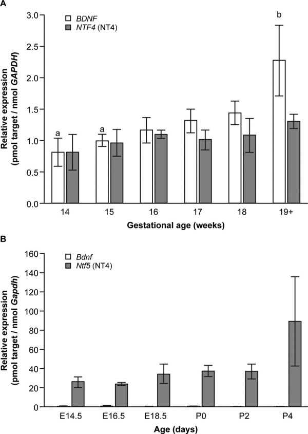
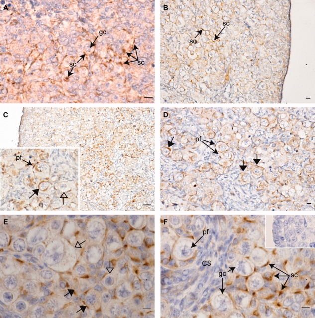
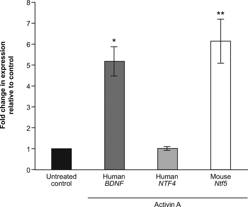
The tropomyosin-related kinase (Trk) B neurotrophin receptor is essential for ovarian germ cell survival and primordial follicle formation, but the contributions of its ligands, brain-derived neurotrophic factor (BDNF) and neurotrophin-4 (NT4), are unknown. We have investigated their expression and regulation in developing human and mouse ovaries. BDNF expression increased with increasing gestation, expression of human NTF4 and of both Ntf5 and Bdnf in the mouse was unchanged. Bdnf expression was dramatically lower than Ntf5 in the mouse, but levels were comparable in the human. Human fetal ovarian somatic cells expressed BDNF. Activin A selectively regulated BDNF and Ntf5 expression in human and mouse, respectively, identifying an oocyte/somatic signaling pathway which might mediate the pro-survival effects of activin. These data reveal that expression and regulation of the TrkB ligands are differentially controlled in the developing ovaries of humans and mice, and identify BDNF as a potential regulator of germ cell fate in the human fetal ovary.


Figures

Fig. 1
Fig. 1
Expression of the genes encoding brain-derived neurotrophic factor (BDNF) and neurotrophin-4 (NT4) in the developing human and mouse ovary. A: Expression of BDNF and NTF4 in the human fetal ovary. Expression of BDNF increased with gestation, and was significantly higher around the time of primordial follicle formation than at early gestations (a vs. b; P < 0.03). In contrast to the mouse, BDNF expression was comparable to that of NTF4. Expression of NTF4 did not change significantly over gestation. B: Expression of Bdnf and Ntf5 in the mouse feto-neonatal ovary from the onset of meiosis (embryonic day [E] 14.5) to primordial follicle formation (postnatal day [P] 2). Ntf5 transcript levels rose slightly with increasing gestation, peaking around the onset of primordial follicle formation, but this was not significant. Bdnf levels were consistently very low at all gestations examined and showed no change over the period examined.
Fig. 2
Fig. 2
Immunohistochemical localization of brain-derived neurotrophic factor (BDNF) in the developing human fetal ovary. BDNF expression was detected at all stages examined. A: Expression of BDNF (brown staining) in somatic cells (sc) surrounding primordial germ cells (gc) in a first trimester (60 days gestation) human fetal ovary. B,C: At 14 (B) weeks and 20 weeks (C); BDNF is predominantly expressed in a corticomedullary gradient, with weak expression in somatic cells near the ovarian periphery, and intense expression in those interspersed between larger germ cells away from the periphery. C, inset: BDNF expression is detectable in the pregranulosa cells of primordial follicles (pf), and in some large germ cells (closed arrows), whereas others of comparable size and show no expression (open arrows). D: At 20 weeks; BDNF immunopositive germ cells are detectable within primordial follicles. E: At 18 weeks; BDNF expression varies between germ cells within the same nest, with immunopositive and immunonegative germ cells in existing close proximity. F: At 18 weeks; BDNF expression is strongest in somatic cells interspersed within germ cell nests and in primordial follicles. No expression is detectable in somatic cells within cell streams (cs). F, inset: negative control, primary antibody preincubated with immunizing peptide. Scale bars = 500 μm in A–D, 100 μm in E,F).
Fig. 3
Fig. 3
Selective regulation of tropomyosin-related kinase (Trk) B ligand expression by Activin A in cultures of human fetal ovary and mouse neonatal ovarian somatic cells. Treatment of disaggregated second trimester human fetal ovaries (14–17 weeks gestational age, n = 5) with recombinant human activin A increased brain-derived neurotrophic factor (BDNF) expression five-fold relative to untreated controls (5.2 ± 0.7; P = 0.02), but had no effect on the expression of NTF4 (1.01 ± 0.09, not significant). Comparable treatments of newborn mouse granulosa cells with activin for 5 days resulted in a six-fold (6.1 ± 1.05; P = 0.01; n = 4) increase in Ntf5 gene expression. Bdnf transcript levels were too low to be reliably determined both in controls and after activin treatment.

References

    1. Anderson RA, Robinson LL, Brooks J, Spears N. Neurotropins and their receptors are expressed in the human fetal ovary. J Clin Endocrinol Metab. 2002;87:890–897. - PubMed
    1. Baker TG. A quantitative and cytological study of germ cells in human ovaries. Proc R Soc Lond B Biol Sci. 1963;158:417–433. - PubMed
    1. Bayne RA, Eddie SL, Collins CS, Childs AJ, Jabbour HN, Anderson RA. Prostaglandin E2 as a regulator of germ cells during ovarian development. J Clin Endocrinol Metab. 2009;94:4053–4060. - PubMed
    1. Bernd P. The role of neurotrophins during early development. Gene Expr. 2008;14:241–250. - PMC - PubMed
    1. Bibel M, Barde YA. Neurotrophins: key regulators of cell fate and cell shape in the vertebrate nervous system. Genes Dev. 2000;14:2919–2937. - PubMed

Publication types

MeSH terms

LinkOut - more resources