

Reduced brain gray matter concentration in patients with obstructive sleep apnea syndrome
- PMID: 20175407
- PMCID: PMC2817910
- DOI: 10.1093/sleep/33.2.235
Reduced brain gray matter concentration in patients with obstructive sleep apnea syndrome
Abstract
Study objectives: To investigate differences in brain gray matter concentrations or volumes in patients with obstructive sleep apnea syndrome (OSA) and healthy volunteers.
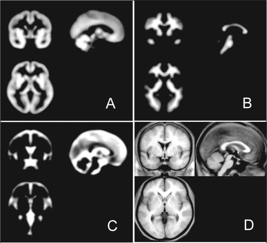
Designs: Optimized voxel-based morphometry, an automated processing technique for MRI, was used to characterize structural differences in gray matter in newly diagnosed male patients.
Setting: University hospital.
Patients and participants: The study consisted of 36 male OSA and 31 non-apneic male healthy volunteers matched for age (mean age, 44.8 years).
Interventions: Using the t-test, gray matter differences were identified. The statistical significance level was set to a false discovery rate P < 0.05 with an extent threshold of k(E) > 200 voxels.
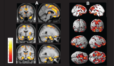
Measurements and results: The mean apnea-hypopnea index (AHI) of patients was 52.5/h. On visual inspection of MRI, no structural abnormalities were observed. Compared to healthy volunteers, the gray matter concentrations of OSA patients were significantly decreased in the left gyrus rectus, bilateral superior frontal gyri, left precentral gyrus, bilateral frontomarginal gyri, bilateral anterior cingulate gyri, right insular gyrus, bilateral caudate nuclei, bilateral thalami, bilateral amygdalo-hippocampi, bilateral inferior temporal gyri, and bilateral quadrangular and biventer lobules in the cerebellum (false discovery rate P < 0.05). Gray matter volume was not different between OSA patients and healthy volunteers.
Conclusions: The brain gray matter deficits may suggest that memory impairment, affective and cardiovascular disturbances, executive dysfunctions, and dysregulation of autonomic and respiratory control frequently found in OSA patients might be related to morphological differences in the brain gray matter areas.
Figures
References
-
- Young T, Peppard PE, Gottlieb DJ. Epidemiology of obstructive sleep apnea: a population health perspective. Am J Respir Crit Care Med. 2002;165:1217–39. - PubMed
-
- Bennett LS, Barbour C, Langford B, Stradling JR, Davies RJ. Health status in obstructive sleep apnea: relationship with sleep fragmentation and daytime sleepiness, and effects of continuous positive airway pressure treatment. Am J Respir Crit Care Med. 1999;159:1884–90. - PubMed
-
- Decary A, Rouleau I, Montplaisir J. Cognitive deficits associated with sleep apnea syndrome: a proposed neuropsychological test battery. Sleep. 2000;23:369–81. - PubMed
-
- Montplaisir J, Bedard MA, Richer F, Rouleau I. Neurobehavioral manifestations in obstructive sleep apnea syndrome before and after treatment with continuous positive airway pressure. Sleep. 1992;15:S17–9. - PubMed
-
- Valencia-Flores M, Bliwise DL, Guilleminault C, Cilveti R, Clerk A. Cognitive function in patients with sleep apnea after acute nocturnal nasal continuous positive airway pressure (CPAP) treatment: sleepiness and hypoxemia effects. J Clin Exp Neuropsychol. 1996;18:197–210. - PubMed
Publication types
MeSH terms
LinkOut - more resources
Full Text Sources
Medical
