. 2010 Mar 30;49(12):2753-62.
doi: 10.1021/bi902200n.

A rearrangement of the guanosine-binding site establishes an extended network of functional interactions in the Tetrahymena group I ribozyme active site

Affiliations

A rearrangement of the guanosine-binding site establishes an extended network of functional interactions in the Tetrahymena group I ribozyme active site

Marcello Forconi et al. Biochemistry. .

Abstract

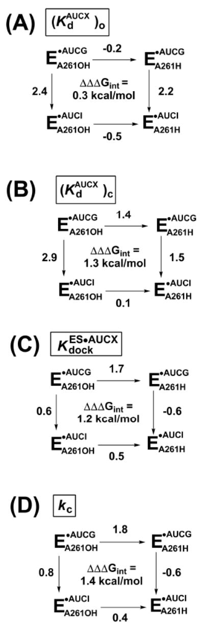
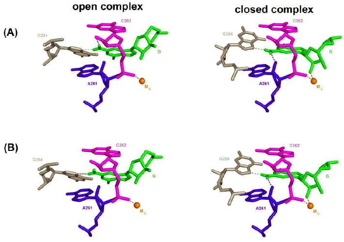
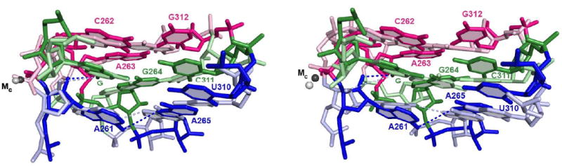
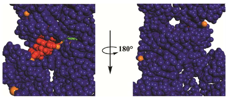
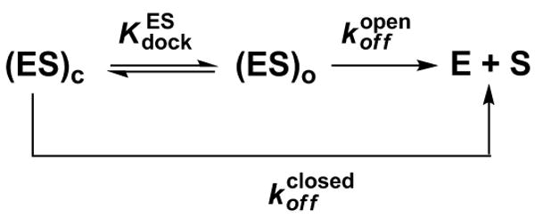
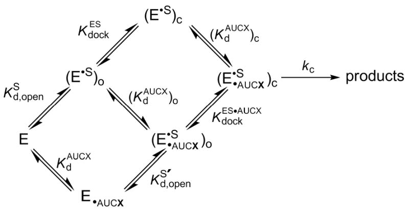
Protein enzymes appear to use extensive packing and hydrogen bonding interactions to precisely position catalytic groups within active sites. Because of their inherent backbone flexibility and limited side chain repertoire, RNA enzymes face additional challenges relative to proteins in precisely positioning substrates and catalytic groups. Here, we use the group I ribozyme to probe the existence, establishment, and functional consequences of an extended network of interactions in an RNA active site. The group I ribozyme catalyzes a site-specific attack of guanosine on an oligonucleotide substrate. We previously determined that the hydrogen bond between the exocyclic amino group of guanosine and the 2'-hydroxyl group at position A261 of the Tetrahymena group I ribozyme contributes to overall catalysis. We now use functional data, aided by double mutant cycles, to probe this hydrogen bond in the individual reaction steps of the catalytic cycle. Our results indicate that this hydrogen bond is not formed upon guanosine binding to the ribozyme but instead forms at a later stage of the catalytic cycle. Formation of this hydrogen bond is correlated with other structural rearrangements in the ribozyme's active site that are promoted by docking of the oligonucleotide substrate into the ribozyme's active site, and disruption of this interaction has deleterious consequences for the chemical transformation within the ternary complex. These results, combined with earlier results, provide insight into the nature of the multiple conformational steps used by the Tetrahymena group I ribozyme to achieve its active structure and reveal an intricate, extended network of interactions that is used to establish catalytic interactions within this RNA's active site.


Figures

FIGURE 1
FIGURE 1. Representation of the transition state of the reaction catalyzed by the Tetrahymena group I ribozyme
The oligonucleotide substrate S (CCCUCUA) is shown on top, and the guanosine nucleophile is below. The metal ion interacting with the deprotonated 3′-OH of the guanosine nucleophile (MB, in grey) is shown as a separate metal ion from that interacting with the 2′-OH of the same residue, as inferred from functional data (39, 64); structural data suggest that the 3′-OH and the 2′-OH of the guanosine nucleophile interact with the same metal ion, MC (grey interaction with the 3′-oxygen of G; refs. (32, 61)). This unresolved issue does not impact the interpretation of the results herein. The hydrogen bond monitored in this work is highlighted by the red box.
FIGURE 2
FIGURE 2. Double-mutant cycles for individual reaction steps
Reactions for WT (EA261OH) or 2′-deoxy A261 (EA261H) ribozyme with AUCG or AUCI. AUCX represents either AUCG or AUCI. The numbers next to each arrow represent the functional effect (ΔΔG in kcal/mol) of either replacing the 2′-hydroxyl group of residue A261 with a 2′-H residue (horizontal arrows) or ablating the exocyclic amine of AUCG, by use of AUCI as the nucleophile (vertical arrows). Values of ΔΔΔGint are calculated by subtracting the value on the right from the value on the right or, equivalently, the value on the bottom from the value on the top. The individual reaction steps (defined in Scheme 2) are as follows: (A) nucleophile binding to the open complex; (B) nucleophile binding to the closed complex; (C) docking of the oligonucleotide substrate S with bound nucleophile; and (D) the chemical step. ΔΔG values are from the rate and equilibrium constants of Tables 2, 3, and 4, and were rounded to a single decimal place to take in account the experimental errors.
FIGURE 3
FIGURE 3. Models for the interactions formed by the exocyclic amino group of AUCG in the open and closed complexes of the WT ribozyme
Representation of the closed complex is from the Azoarcus 3bo3 structural model (41); the model for the open complex is obtained by arbitrarily rotating the G nucleophile and G264. In model (A) the exocyclic amino group of AUCG forms two hydrogen bonds with the ribozyme (represented by dashed lines) in both the open and the closed complex, but one of these hydrogen bonds is different in the two complexes; the hydrogen bond between the exocyclic amino group of G and the 2′-hydroxyl group of residue C262, shown in the open complex, is purely speculative. In contrast, for model (B) only one hydrogen bond is formed with the exocyclic amino group of G264 in the open complex, and is represented by a thicker line. In both models the closed complex is the same with the exocyclic amino group of the G nucleophile making hydrogen bonds with the 2′-hydroxyl group of residue A261 and the N7 atom of G264. The contact with G264 is inferred from previous functional data (31) and suggested from structural data (32-34, 41, 61) but has not been experimentally tested. This and the following figures were generated with Pymol (DeLano, W.L. The PyMOL Molecular Graphics System (2002) on World Wide Web http://www.pymol.org)
FIGURE 4
FIGURE 4. Superpositioning of structural models of the guanosine binding site from different group I introns crystal structures reveal subtle differences
Model from the Azoarcus intron, PDB IS 3bo3 (41), is depicted in darker colors; models from the Tetrahymena intron (34) are in lighter colors and correspond to molecule C from the Tetrahymena ribozyme crystals (part A) and molecule A (part B). Numbering of residues is according to the Tetrahymena intron. Structures were superimposed by aligning the guanosine nucleophile (G). Nucleobases proposed to be part of the same base triple are depicted in the same color. Dashed lines connect the 2′-hydroxyl group of residue A261 and the exocyclic nitrogen atom of G, representing the hydrogen bond functionally detected and investigated herein, and the exocyclic nitrogen atom of residue A261 and the N7 atom of residue A265, representing a hydrogen bond proposed to stabilize the guanosine-binding site. Numerous other proposed hydrogen bonds within this site are omitted for clarity.
FIGURE 5
FIGURE 5. Network of interactions in the group I ribozyme active site
Residues are numbered according to the Tetrahymena intron and shown for the Azoarcus 3bo3 structural model (41). This structure contains the products of the reactions carried out herein, with the reactive phosphoryl group and the A residue transferred on the 3′-oxygen of the guanosine nucleophile. Different regions of the ribozyme are colored differently. The two active site metal ions present in the model are shown as orange and yellow spheres and correspond to MC and MA in Figure 1, respectively. Dashed lines represent metal ion contacts or hydrogen bonds and are color coded according to their location. Atoms involved in these contacts are represented by spheres, blue for nitrogen and red for oxygen; hydrogen atoms are now shown. The hydrogen bond between the 2′-hydroxyl group of residue A261 and the exocyclic amino group of G, investigated herein, is represented by the dashed blue line. Stacking of the nucleobases of residues C262, A261, A306 and the G nucleophile is suggested from the X-ray structures (32-34, 41, 61). Additional base triples and stacking interactions are omitted for clarity (see Figure 4).
FIGURE 6
FIGURE 6. Space-filling representation of the environment around the guanosine nucleophile
The guanosine nucleophile is in green, the oligonucleotide substrate in red, metal ions in orange, and the ribozyme in blue. The guanosine nucleophile is shown as a stick representation and all other atoms as space filling. Only the central portion of the Azoarcus structural model is shown (3bo3, ref. 41).
SCHEME 1
SCHEME 1
SCHEME 2
SCHEME 2

Similar articles

Cited by

References

    1. Jencks WP. Economics of enzyme catalysis. Cold Spring Harb Symp Quant Biol. 1987;52:65–73. - PubMed
    1. Ditzler MA, Aleman EA, Rueda D, Walter NG. Focus on function: single molecule RNA enzymology. Biopolymers. 2007;87:302–316. - PubMed
    1. Johnson TH, Tijerina P, Chadee AB, Herschlag D, Russell R. Structural specificity conferred by a group I RNA peripheral element. Proc Natl Acad Sci USA. 2005;102:10176–10181. - PMC - PubMed
    1. Xie Z, Srividya N, Sosnick TR, Pan T, Scherer NF. Single-molecule studies highlight conformational heterogeneity in the early folding steps of a large ribozyme. Proc Natl Acad Sci USA. 2004;101:534–539. - PMC - PubMed
    1. Blount KF, Uhlenbeck OC. The structure-function dilemma of the hammerhead ribozyme. Annu Rev Biophys Biomol Struct. 2005;34:415–440. - PubMed

Publication types