. 2010 Jul 1;64(1):27-37.
doi: 10.1111/j.1600-0897.2010.00817.x. Epub 2010 Feb 17.

Viral ssRNA induces first trimester trophoblast apoptosis through an inflammatory mechanism

Affiliations

Viral ssRNA induces first trimester trophoblast apoptosis through an inflammatory mechanism

Paulomi B Aldo et al. Am J Reprod Immunol. .

Abstract

Problem: Infection during pregnancy represents a significant cause of mobility and mortality. While viruses pose a major threat, little is known about their effect on early pregnancy, or the mechanisms involved. The objective of this study was to characterize the trophoblast response following exposure to viral ssRNA.

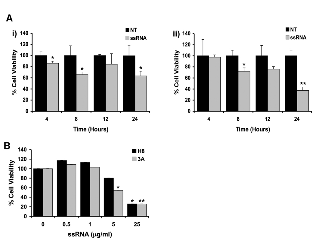
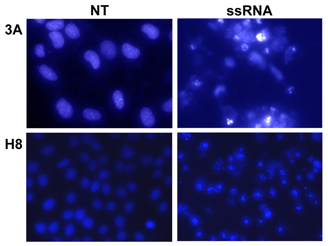
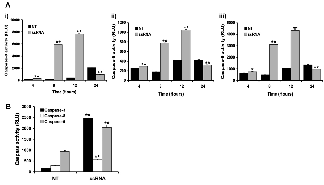
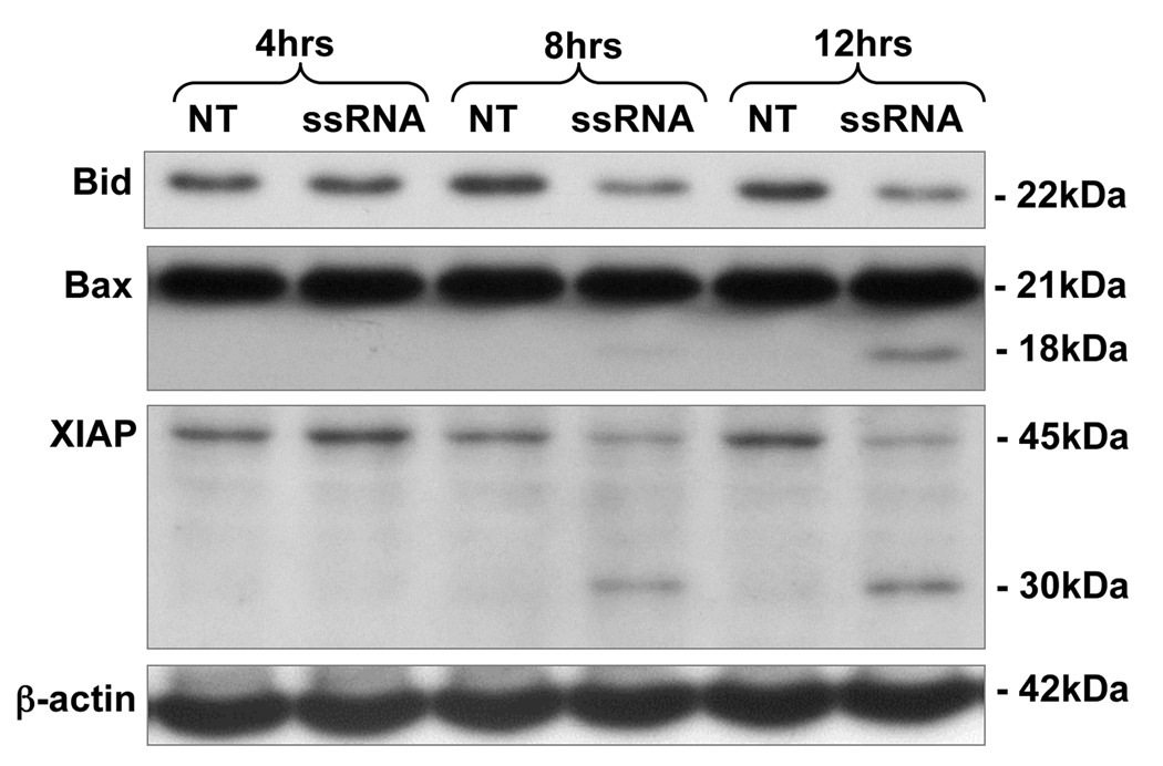
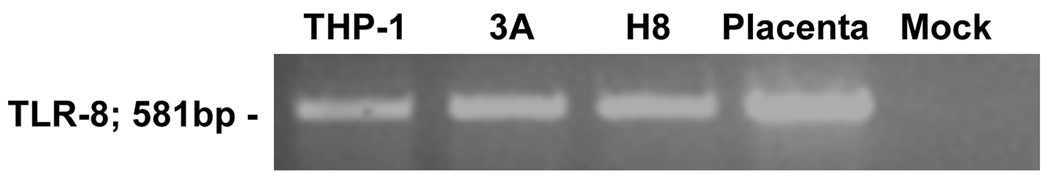
Method of study: First trimester trophoblast cells were treated with or without viral ssRNA. Cytokine production was measured using multiplex analysis and ELISA. Apoptosis was determined using Hoechst staining, cell viability, and caspase activity assays.

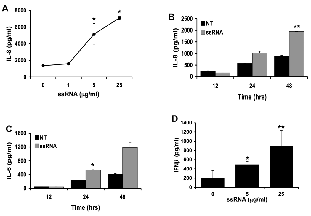
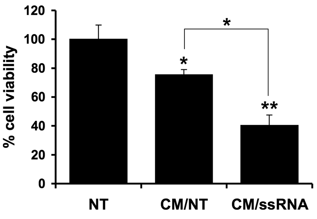
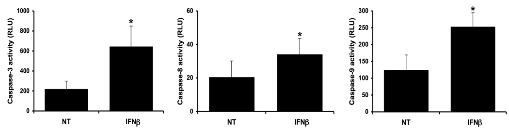
Results: Treatment of trophoblasts with viral ssRNA increased their secretion of IL-8, IL-6, and IFNbeta. However, the ssRNA also induced trophoblast apoptosis. To test whether the viral ssRNA-induced inflammatory response was responsible for this induction of apoptosis, conditioned media (CM) from trophoblasts were added to a fresh culture of cells. The CM from viral ssRNA-treated induced higher levels of trophoblast apoptosis than the control CM. Moreover, recombinant IFNbeta induced trophoblast apoptosis.

Conclusion: We demonstrate that viral ssRNA induces a pro-inflammatory and type I interferon response in the trophoblast and this inflammatory process may indirectly induce trophoblast apoptosis. These results provide a novel mechanism by which certain viral infections might compromise placental integrity and function, and therefore, pregnancy outcome.


Figures

Figure 1
Figure 1. Viral ssRNA reduces first trimester trophoblast cell viability
(A) The first trimester trophoblast cells, (i) H8 and (ii) 3A, were incubated with either no treatment (NT), or ssRNA (25⎧g/ml) for 4, 8, 12 and 24 hours. (B) The first trimester trophoblast cells, H8 and 3A, were incubated with viral ssRNA at 0, 0.5, 1, 5 or 25⎧g/ml for 48 hours. Following each time point, cell viability was determined using the CellTiter 96 assay. Barcharts show percentage cell viability relative to the untreated control. Treatment with ssRNA significantly reduced trophoblast cell viability in a dose and time dependent manner (*p <0.05, **p <0.001). Data are representative of at least three independent experiments.
Figure 2
Figure 2. Viral ssRNA induces apoptosis in first trimester trophoblast cells
The first trimester trophoblast cells, 3A (Mag. 40×) and H8 (Mag. 20×) were incubated for 24 hours with either no treatment (NT) or ssRNA (25⎧g/ml). Following incubation, the cells were stained with Hoechst 33342 dye (5⎧g/ml) and cells visualized by fluorescent microscopy. Note the increased numbers of condensed and fragmented nuclei in the ssRNA treated cells. Data are representative of at least three independent experiments.
Figure 3
Figure 3. Viral ssRNA activates the apoptotic caspase pathway in first trimester trophoblast cells
(A) The first trimester trophoblast cells, 3A, were incubated with either no treatment (NT) or ssRNA (25⎧g/ml) for 4, 8, 12 or 24 hours, after which cell lysates were prepared and: (i) caspase-3; (ii) caspase-8; and (iii) caspase-9 activities were determined using the Caspase-Glo assay. (B) H8 cells were incubated with either no treatment (NT) or ssRNA (25⎧g/ml) for 12 hours, after which caspase-3, caspase-8 and caspase-9 activities were determined. Bar charts show caspase activity in relative light units (RLU). Viral ssRNA significantly increased trophoblast cell caspase-3, caspase-8 and capsase-9 activity, with levels peaking after 12 hours of treatment. *p<0.05; **p<0.001 relative to the NT control. Data are representative of at least three independent experiments.
Figure 4
Figure 4. Viral ssRNA alters apoptotic protein expression in first trimester trophoblast cells
First trimester trophoblast cells (3A) were incubated with either no treatment (NT) or ssRNA (25⎧g/ml) for 4, 8 or 12 hours. After each time point, cell lysates were prepared and analyzed for Bid, Bax and XIAP expression levels by Western blot. ®-actin served as a loading control. Data are representative of three independent experiments.
Figure 5
Figure 5. First trimester trophoblast cells express Toll-like receptor 8
TLR-8 mRNA expression was evaluated in first trimester trophoblast cells by RT-PCR. Panel shows TLR-8 expression (581bp) in the 3A and H8 trophoblast cells as well as 8-week placental tissue. The monocytic THP-1 cell line served as a positive control. No signal was detected in the mock control.
Figure 6
Figure 6. Viral ssRNA triggers an inflammatory cytokine response in trophoblast cells
First trimester trophoblast cells, (H8), were incubated with either: (A) ssRNA at 0, 1, 5 or 25⎧g/ml for 48 hours; (B&C) no treatment (NT) or ssRNA (5⎧g/ml) for 12, 24 or 48 hours; or (D) ssRNA at 0, 5 or 25⎧g/ml for 48 hours. Cell-free supernatants were collected and analyzed for IL-8 and IL-6 secretion by multiplex analysis, and IFN® secretion by ELISA. *p<0.05; **p<0.001 relative to the untreated control. Data are representative of at least three independent experiments.
Figure 7
Figure 7. Effect of viral-ssRNA induced trophoblast-derived factors on trophoblast cell viability
First trimester trophoblast cells (H8) were treated with no treatment (NT) or ssRNA (5⎧g/ml) for 48 hours, after which the cell-free conditioned media (CM) was collected. A fresh culture of H8 cells were then incubated with either: no treatment (NT); 50% of CM from untreated trophoblast cells (CM/NT); or 50% of CM from trophoblast cells treated with ssRNA (CM/ssRNA). After 96 hours, cell viability was determined using the CellTiter™ assay. *p<0.05 and **p<0.001 relative to the NT control, unless otherwise indicated. Data are representative of at least three independent experiments.
Figure 8
Figure 8. IFN® induces trophoblast apoptosis
First trimester trophoblast cells (H8) were treated with no treatment (NT) or recombinant human IFN® (10,000U/ml) for 72 hours, after which (i) caspase-3; (ii) caspase-8; and (iii) caspase-9 activities were determined. *p<0.001 relative to the NT control. Data are from four independent experiments.

Similar articles

Cited by

References

    1. Espinoza J, Erez O, Romero R. Preconceptional antibiotic treatment to prevent preterm birth in women with a previous preterm delivery. Am J Obstet Gynecol. 2006;194:630–637. - PubMed
    1. Pereira L, Maidji E, McDonagh S, Tabata T. Insights into viral transmission at the uterine-placental interface. Trends Microbiol. 2005;13:164–174. - PubMed
    1. Goldenberg RL, Hauth JC, Andrews WW. Intrauterine infection and preterm delivery. N Engl J Med. 2000;342:1500–1507. - PubMed
    1. Lamont RF. The role of infection in preterm labour and birth. Hosp Med. 2003;64:644–647. - PubMed
    1. Arechavaleta-Velasco F, Koi H, Strauss JF, 3rd, Parry S. Viral infection of the trophoblast: time to take a serious look at its role in abnormal implantation and placentation? J Reprod Immunol. 2002;55:113–121. - PubMed

Publication types