. 2010 Apr;23(2):276-86.
doi: 10.1111/j.1755-148X.2010.00688.x. Epub 2010 Feb 19.

Reduced skin homing by functional Treg in vitiligo

Affiliations

Reduced skin homing by functional Treg in vitiligo

Jared Klarquist et al. Pigment Cell Melanoma Res. 2010 Apr.

Erratum in

  • Pigment Cell Melanoma Res. 2010 Jun;23(3):477. Wainwright, Derek J [corrected to Wainwright, Derek A]

Abstract

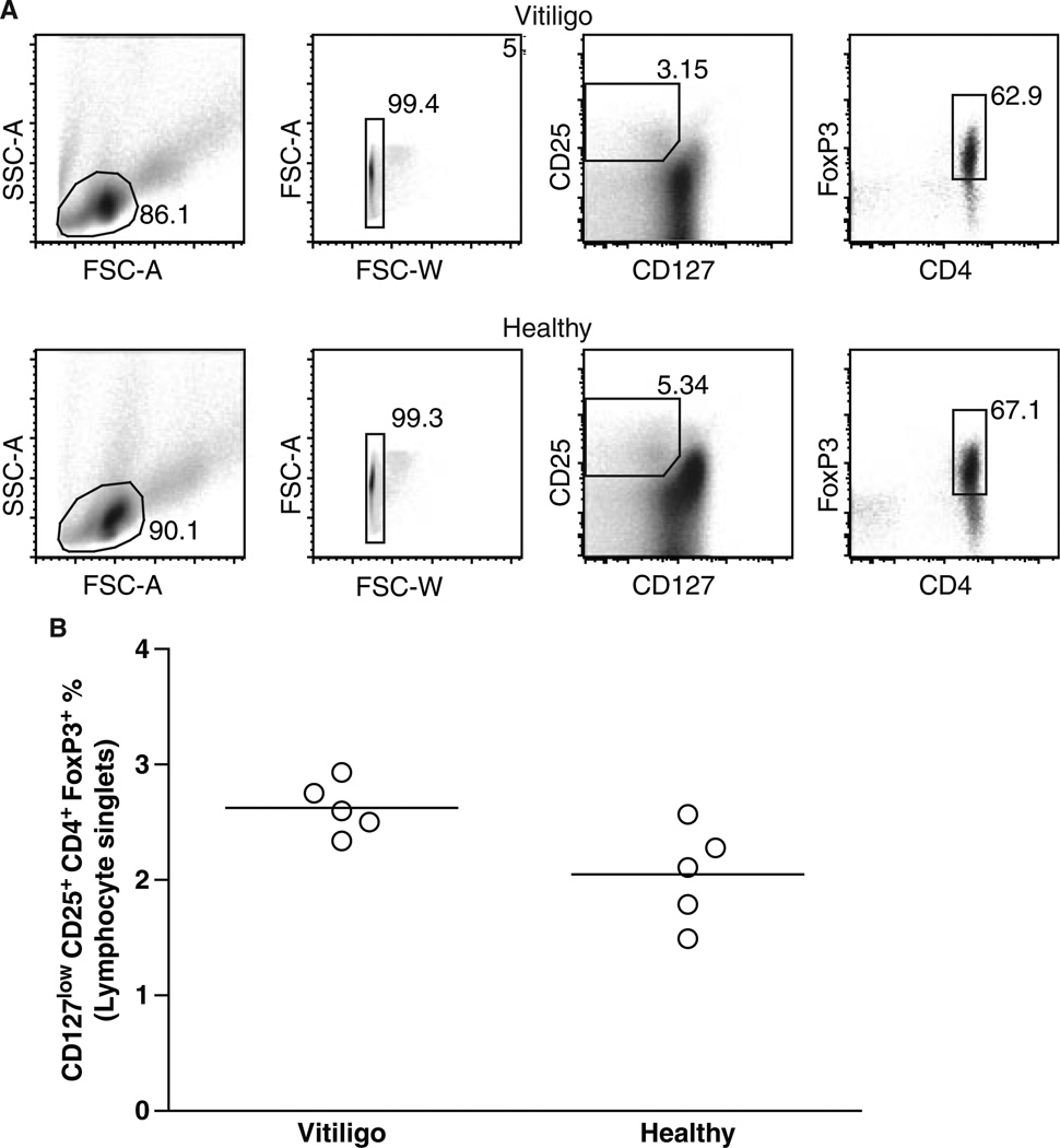
In human vitiligo, cutaneous depigmentation involves cytotoxic activity of autoreactive T cells. It was hypothesized that depigmentation can progress in the absence of regulatory T cells (Treg). The percentage of Treg among skin infiltrating T cells was evaluated by immunoenzymatic double staining for CD3 and FoxP3, revealing drastically reduced numbers of Treg in non-lesional, perilesional and lesional vitiligo skin. Assessment of the circulating Treg pool by FACS analysis of CD4, CD25, CD127 and FoxP3 expression, and mixed lymphocyte reactions in presence and absence of sorted Treg revealed no systemic drop in the abundance or activity of Treg in vitiligo patients. Expression of skin homing receptors CCR4, CCR5, CCR8 and CLA was comparable among circulating vitiligo and control Treg. Treg from either source were equally capable of migrating towards CCR4 ligand and skin homing chemokine CCL22, yet significantly reduced expression of CCL22 in vitiligo skin observed by immunohistochemistry may explain failure of circulating, functional Treg to home to the skin in vitiligo. The paucity of Treg in vitiligo skin is likely crucial for perpetual anti-melanocyte reactivity in progressive disease.


Figures

Figure 1
Figure 1
Paucity of Treg in vitiligo skin. Treg abundance quantified. (A) Immunostaining of all T cells (blue) and Tregs (red) was light microscopically evaluated by two investigators. (B) The percent Treg among T cells was calculated for 4–8 samples in each group, looking for significant differences to the % Treg found in adult normal skin by Student’s T test, with **P < 0.01, and *P < 0.05.
Figure 2
Figure 2
Abundance of circulating Treg quantified by FACS analysis. (A) Representative FACS plots for a vitiligo and a control blood sample, with CD4+CD25+CD127low and FoxP3+ used to identify Treg. (B) The average percentage of Treg among lymphocytes was quantified and compared among control and vitiligo samples. According to these data, the number of Treg circulating in patients does not support a systemic defect in Treg in vitiligo.
Figure 3
Figure 3
Inhibition of T cell proliferation quantified. Proliferation of CD4+CD25 lymphocytes from patients and controls was quantified by 3H-thymidine incorporation in the presence and absence of the CD25+ subpopulation of CD4+ cells and allogeneic DC to stimulate mixed lymphocyte responses, showing that circulating Treg from vitiligo patients (n = 3) are equally capable of immunosuppression as Treg from control individuals (n = 3). The data further support that Treg from vitiligo patients do not display a systemic defect to explain their paucity in vitiligo skin.
Figure 4
Figure 4
Skin homing marker expression among circulating Treg analyzed by FACS. (A) Representative control subject plots show sequential gating strategies for identifying Treg, as well as CD4+FoxP3 and CD4FoxP3 subsets among CD3+ enriched lymphocytes. These subsets were analyzed for expression of CCR4, CCR5, CCR8 and CLA. Summarized data (B) show a similar percentage of skin homing marker expression among patient and control Treg, and CD4FoxP3 populations whereas CCR8 expression among non-Treg T cells was significantly higher in vitiligo subjects (not shown). The MFI representing CCR4, CCR5, CCR8 and CLA expression was similar among patient and control Treg.
Figure 5
Figure 5
Skin homing chemokine CCL22 is markedly reduced in vitligo patient skin. Representative images are given for control (A) and non-lesional vitiligo (B) frozen skin immunostained for CCL22. (C) The data reveal a significantly reduced abundance of CCL22 expressing non-lesional and intra-lesional vitiligo skin (P < 0.05). Treg migration in response to CCL22 was also determined in control and vitiligo samples (D). Treg abundance among migrated CD4+ lymphocytes was measured in the absence (spontaneous migration) or presence of CCR4 ligand CCL22. Treg migration data were summarized showing no difference in the % Treg responding to CCL22 among control and vitiligo CD4+ lymphocytes.

References

    1. Al-Mutairi N, Al-Doukhi A. Familial coexisting and colocalized psoriasis and vitiligo responding to alefacept. J. Cutan. Med. Surg. 2009;13:172–175. - PubMed
    1. Baecher-Allen C, Hafler DA. Human regulatory T cells and their role on autoimmune disease. Immunol. Rev. 2006;212:203–216. - PubMed
    1. Bala KK, Moudgil KD. Induction and maintenance of self tolerance: the role of CD4+CD25+ regulatory T cells. Arch. Immunol. Ther. Exp. (Warsz) 2006;54:307–321. - PubMed
    1. Chakraborty NG, Chattopadhyay S, Mehrotra S, Chhabra A, Mukherji B. Regulatory T-cell reponse and tumor vaccine-induced cytotoxic T lymphocytes in human melanoma. Hum. Immunol. 2004;65:794–802. - PubMed
    1. Colantonio L, Iellem A, Sinigaglia F, D’Ambrosio D. Skin-homing CLA+ T cells and regulatory CD25+ T cells represent major subsets of human peripheral blood memory T cells migrating in response to CCL1 / I-309. Eur. J. Immunol. 2002;32:3506–3514. - PubMed