Case Reports
. 2010 Feb 22:2:5.
doi: 10.1186/1758-3284-2-5.

Establishment and characterization of a novel head and neck squamous cell carcinoma cell line USC-HN1

Affiliations
Case Reports

Establishment and characterization of a novel head and neck squamous cell carcinoma cell line USC-HN1

Daniel J Liebertz et al. Head Neck Oncol. .

Abstract

Background: Head and neck squamous cell carcinoma (HNSCC) is an aggressive and lethal malignancy. Publically available cell lines are mostly of lingual origin, or have not been carefully characterized. Detailed characterization of novel HNSCC cell lines is needed in order to provide researchers a concrete keystone on which to build their investigations.

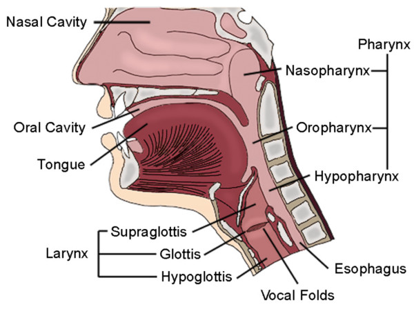
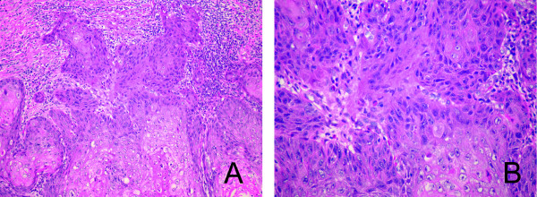
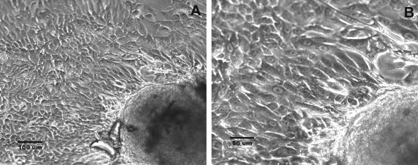
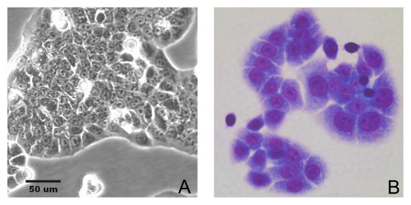
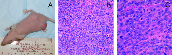
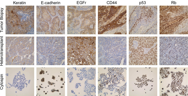
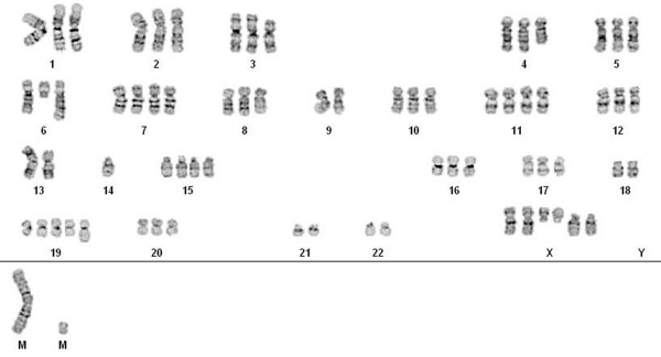
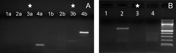
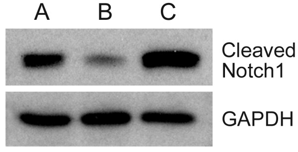
Methods: The USC-HN1 cell line was established from a primary maxillary HNSCC biopsy explant in tissue culture. The immortalized cells were then further characterized by heterotransplantation in Nude mice; immunohistochemical staining for relevant HNSCC biomarkers; flow cytometry for surface markers; cytogenetic karyotypic analysis; human papillomavirus and Epstein-Barr virus screening; qRT-PCR for oncogene and cytokine analysis; investigation of activated, cleaved Notch1 levels; and detailed 35,000 gene microarray analysis.

Results: Characterization experiments confirmed the human HNSCC origin of USC-HN1, including a phenotype similar to the original tumor. Viral screening revealed no HPV or EBV infection, while western blotting displayed significant upregulation of activated, cleaved Notch1.

Conclusions: USC-HN1, a novel immortalized cell line has been derived from a maxillary HNSCC. Characterization studies have shown that the cell line is of HNSCC origin and displays many of the same markers previously reported in the literature. USC-HN1 is available for public research and will further the investigation of HNSCC and the development of new therapeutic modalities.


Figures

Figure 1
Figure 1
Schematic of the upper aerodigestive tract and locations of various head and neck malignancies.
Figure 2
Figure 2
Hematoxilin and eosin stained histologic sections from the original tumor. An invasive well-differentiated squamous cell carcinoma, the tumor is arranged as cohesive islands of cells showing nuclear pleomorphism with occasionally prominent nucleoli. Mitotic activity is present including atypical forms. In areas, abundant keratin production is present including whirling "keratin pearl" configurations. Intercellular bridges are visible at high power magnification. Surrounding the invasive tumor, a dense lymphoplasmacytic inflammatory infiltrate is present (A ×200, B ×400 original magnification).
Figure 3
Figure 3
Morphologic appearance of early tumor explants from HNSCC biopsy samples. Phase-contrast photomicrograph of tumor explants isolated from HNSCC biopsies in culture after 2-4 weeks with cells growing directly from the explants in a monolayer (A ×100, B ×200 original magnification).
Figure 4
Figure 4
Establishment of the USC-HN1 cell line. (A) Phase contrast microphotograph of growing USC-HN1 cells showing numerous mitotic cells (rounded, luminescent cells) with a tightly, adherent squamous cell morphology. (B) Cytology of the USC-HN1 cell line shows malignant cells with large nuclei and nucleoli and an abundance of cytoplasm typical of squamous cells (Cytospin, Wright-Giemsa stain, ×100 original magnification).
Figure 5
Figure 5
Heterotransplantation of USC-HN1 cell line into Nude mice. (A) Appearance of subcutaneous USC-HN1 tumor in Nude mouse. (B and C) Low and high magnification of USC-HN1 Nude mouse heterotransplant showing a poorly differentiated squamous cell carcinoma arranged as a sheet with areas of tumor necrosis and bluntly infiltrative borders. The tumor cells are tightly cohesive featuring from moderate to abundant eosinophilic cytoplasm. The nuclear to cytoplasmic ratio is markedly increased with nuclei showing frequent, prominent nucleoli. Mitotic activity is abundant including atypical forms such as ring and tripolar configurations. Intercellular bridges are focally present but faint (H&E ×200 and ×400 original magnification).
Figure 6
Figure 6
Immunoperoxidase staining of USC-HN1 cells in Nude mouse heterotransplant and in cytospin preparations for HNSCC classification markers. Photomicrograph of immunoperoxidase staining of original tumor biopsy (top), IHC stained formalin-fixed paraffin-embedded tissue sections of USC-HN1 Nude mouse heterotransplant (middle), and USC-HN1 cells from culture in a cytospin preparation (bottom) for keratin, E-cadherin, EGFr, CD44, p53 and Rb (×200 original magnification).
Figure 7
Figure 7
Karyotype of USC-HN1. Karyotype of USC-HN1 cell line showing a near-triploid clone (modal 71) demonstrating features characteristic of head and neck cancer including multiple deletions (chromosomes X, 3, 4, 6, and 7), isochromosome formation, and many breakpoints around centromeres.
Figure 8
Figure 8
PCR studies for the presence of oncogeneic viruses. (A) HPV detection using consensus primers GP5+/GP6+ (1a-4a) and MY09/MY11 (1b-4b) demonstrating USC-HN1 (3a, 3b) is negative for HPV infection. Positive (HeLa 4a, 4b), negative (SW579 2a, 2b), and water (1a, 1b) controls were run in parallel. (B) PCR study for detection of EBV with consensus primer showing USC-HN1 negative (3). Positive (Raji, 2), negative (HUT102, 4), and water (1) controls were run in parallel.
Figure 9
Figure 9
Western Blot: Notch1. Western blot analysis of the active, cleaved portion of Notch1 run in parallel with confirmatory GAPDH. (lane A) Karpas 299 positive control; (lane B) Siha low expression control; and (lane C) USC-HN1 shows high expression levels of Notch1 protein.

Similar articles

Cited by

References

    1. Lin CJ, Grandis JR, Carey TE, Gollin SM, Whiteside TL, Koch WM, Ferris RL, Lai SY. Head and neck squamous cell carcinoma cell lines: established models and rationale for selection. Head Neck. 2007;29:163–88. doi: 10.1002/hed.20478. - DOI - PubMed
    1. Chiou SH, Yu CC, Huang CY, Lin SC, Liu CJ, Tsai TH, Chou SH, Chien CS, Ku HH, Lo JF. Positive correlations of Oct-4 and Nanog in oral cancer stem-like cells and high-grade oral squamous cell carcinoma. Clin Cancer Res. 2008;14(13):4085–95. doi: 10.1158/1078-0432.CCR-07-4404. - DOI - PubMed
    1. Bergmann C, Strauss L, Wang Y, Szczepanski MJ, Lang S, Johnson JT, Whiteside TL. T regulatory Type 1 cells in squamous cell carcinoma of the head and neck: mechanisms of suppression and expansion in advanced disease. Clin Cancer Res. 2008;14(12):3706–15. doi: 10.1158/1078-0432.CCR-07-5126. - DOI - PMC - PubMed
    1. Gold KA, Kim ES. Role of molecular markers and gene profiling in head and neck cancers. Curr Opin Oncol. 2009;21:206–11. doi: 10.1097/CCO.0b013e328329ac00. - DOI - PubMed
    1. Culliney B, Birhan A, Young AV, Choi W, Shulimovich M, Blum RH. Management of locally advanced or unresectable head and neck cancer. Oncology. 2008;22(10):1152–61. discussion 1162-6, 1171-2. - PubMed

Publication types