

Primary carcinoid tumor of the gallbladder: a case report and brief review of the literature
- PMID: 20175936
- PMCID: PMC2834672
- DOI: 10.1186/1477-7819-8-12
Primary carcinoid tumor of the gallbladder: a case report and brief review of the literature
Abstract
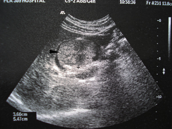
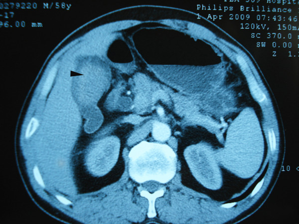
Background: Primary carcinoid tumor of the gallbladder is rare and comprises less than 1% of all carcinoid tumors. Preoperative diagnosis of carcinoid tumor of the gallbladder is difficult. The imageology findings are similar to those in other gallbladder cancers.
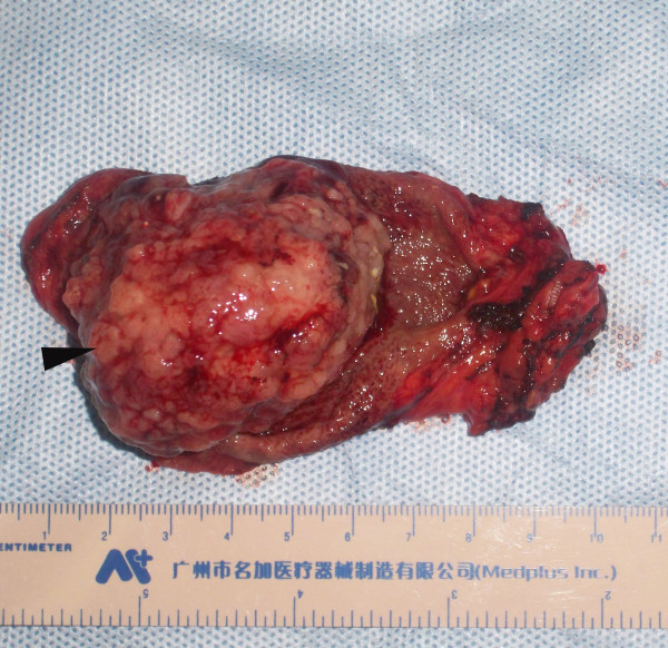
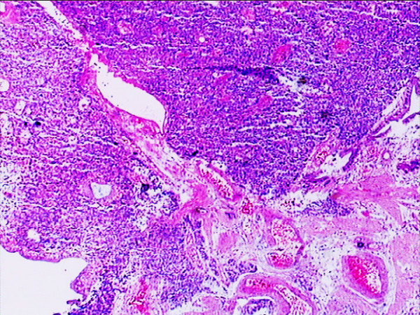
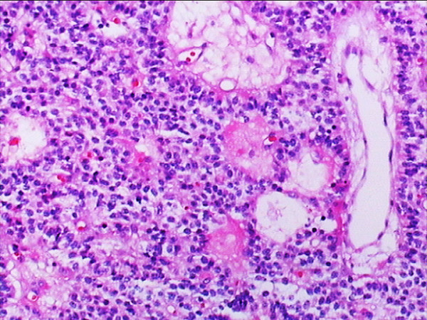
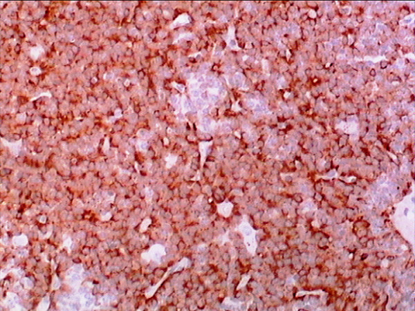
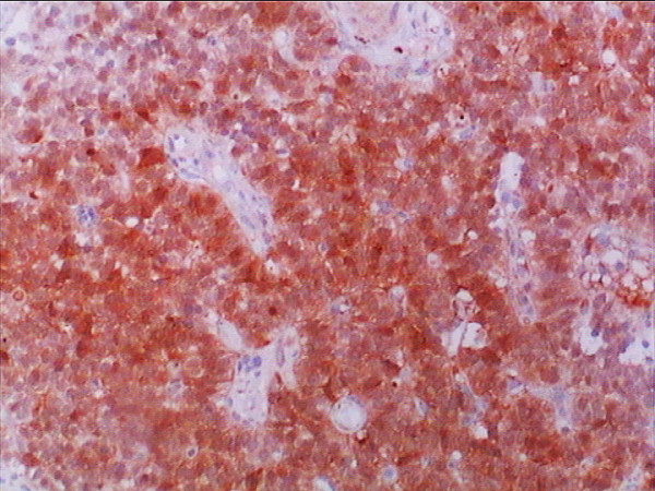
Case presentation: A 46-year-old woman was hospitalized with a preoperative diagnosis of gallbladder carcinoma, The patient was referred for surgical opinion and laparotomy was subsequently performed. A 4 x 5 cm mass was found within the gallbladder, located on the free surface of the body and fundus of the gallbladder. Neither metastases nor direct invasion to the liver was found. The entire mass and gallbladder were excised and intact. Histologically, the tumor consisted of small oval cells with round-to-oval neclei and tumor cells formed small nodular, trabeculare and acinar structures. The tumor showed moderate pleomorphism with scattered mitotic figures, but no definite evidence of vascular permeation, perineural invasion or lymphatic permeation was seen. The tumor cells invaded the mucosa extensively, and some penetrated the muscular layer but not through the serosa of the gallbladder into the liver. Immunohistochemical studies revealed strong positive reaction for chromogranin A and NSE. This lesion was proved to be a primary carcinoid tumor of the gallbladder. A brief review of literature, clinical feature, pathology and treatment of this rare disease was discussed.
Conclusion: Primary carcinoid tumor of the gallbladder is uncommon. The definite diagnosis is often made on histopathological results after surgery.
Figures
Similar articles
-
Carcinoid tumor of the gallbladder: laparoscopic resection and review of the literature.Surgery. 1992 Jul;112(1):100-5. Surgery. 1992. PMID: 1535733 Review.
-
Carcinoid tumor of the gall bladder.Ann Diagn Pathol. 2007 Apr;11(2):113-6. doi: 10.1016/j.anndiagpath.2005.12.003. Ann Diagn Pathol. 2007. PMID: 17349570
-
Gallbladder carcinoid masquerading as gallbladder carcinoma.Hepatobiliary Pancreat Dis Int. 2009 Jun;8(3):326-8. Hepatobiliary Pancreat Dis Int. 2009. PMID: 19502178
-
Carcinoid tumor of the gall bladder.Intern Med. 1996 Dec;35(12):953-6. doi: 10.2169/internalmedicine.35.953. Intern Med. 1996. PMID: 9030993
-
Primary carcinoid tumor of the gallbladder: resection of a case metastasizing to the liver and analysis of outcomes.Hepatogastroenterology. 2000 Jan-Feb;47(31):135-9. Hepatogastroenterology. 2000. PMID: 10690596 Review.
Cited by
-
Polypoid gallbladder neuroendocrine tumor diagnosed as benign polyp before surgery: A case report.Mol Clin Oncol. 2020 Mar;12(3):225-229. doi: 10.3892/mco.2019.1971. Epub 2019 Dec 24. Mol Clin Oncol. 2020. PMID: 32064098 Free PMC article.
-
Small Cell Carcinoma of the Gallbladder.Cureus. 2023 May 2;15(5):e38444. doi: 10.7759/cureus.38444. eCollection 2023 May. Cureus. 2023. PMID: 37273321 Free PMC article.
-
Neuroendocrine Carcinoma of the Gallbladder Masquerading as a Klatskin Tumor in a 74-Year-Old Male.J Gastrointest Cancer. 2016 Mar;47(1):118-22. doi: 10.1007/s12029-015-9713-4. J Gastrointest Cancer. 2016. PMID: 26036328 No abstract available.
-
Gallbladder Cancer in the 21st Century.J Oncol. 2015;2015:967472. doi: 10.1155/2015/967472. Epub 2015 Sep 1. J Oncol. 2015. PMID: 26421012 Free PMC article. Review.
-
Carcinoid tumor of the gallbladder: report of two cases.Clin J Gastroenterol. 2011 Oct;4(5):323-330. doi: 10.1007/s12328-011-0242-9. Epub 2011 Jul 23. Clin J Gastroenterol. 2011. PMID: 26189633
References
-
- Sanders RJ. Carcinoid of the Gastrointestinal Tract. Vol. 10. Springfield, Charles C Thomas Publisher; 1973.
-
- Joel W. Karzinoidtumor der Gallenblase. Zentralbl Allg Pathol. 1929;46:14.
Publication types
MeSH terms
LinkOut - more resources
Full Text Sources
Medical
Research Materials
