

Expression of hyaluronan and the hyaluronan-binding proteoglycans neurocan, aggrecan, and versican by neural stem cells and neural cells derived from embryonic stem cells
- PMID: 20176001
- PMCID: PMC2878141
- DOI: 10.1016/j.brainres.2010.02.048
Expression of hyaluronan and the hyaluronan-binding proteoglycans neurocan, aggrecan, and versican by neural stem cells and neural cells derived from embryonic stem cells
Abstract
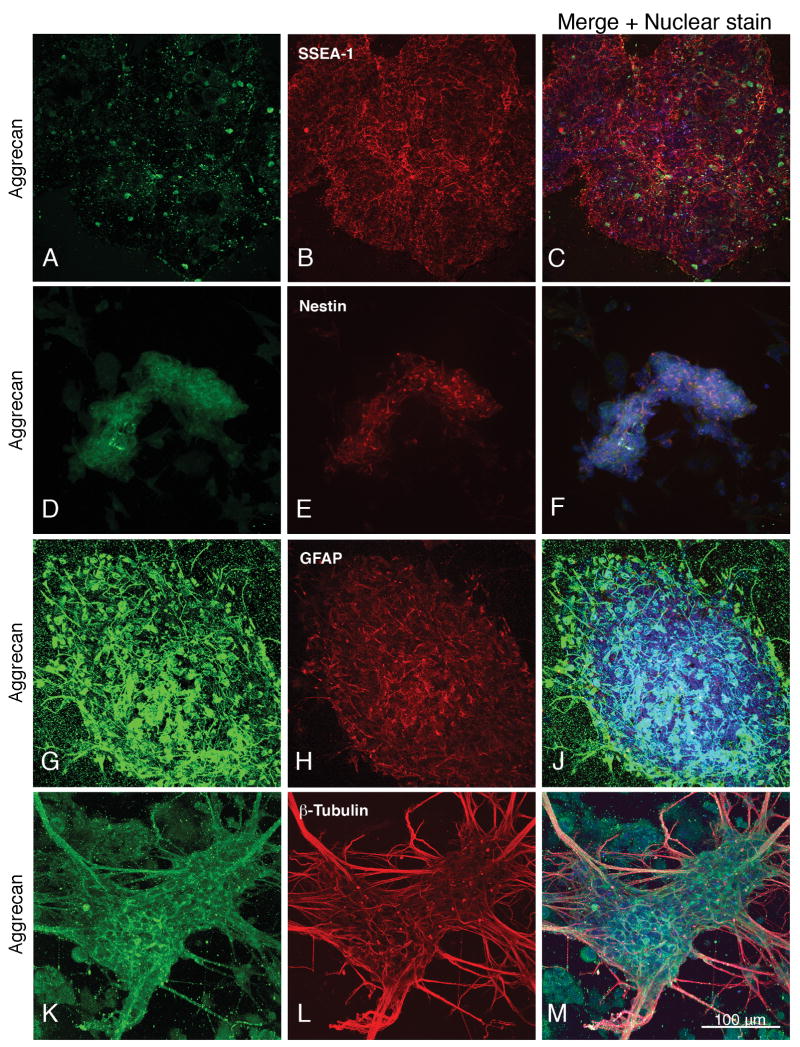
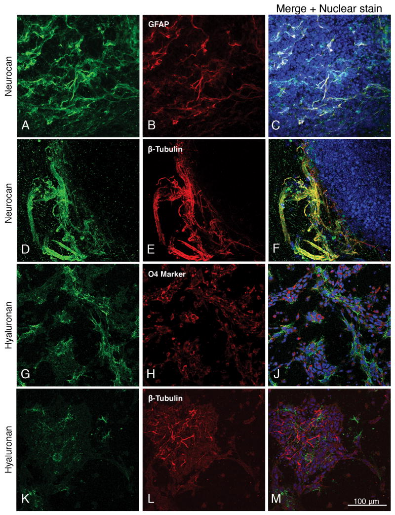
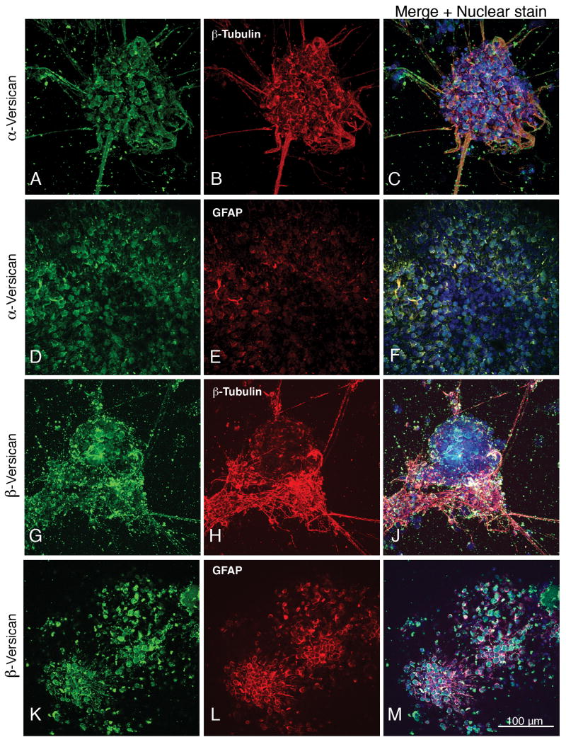
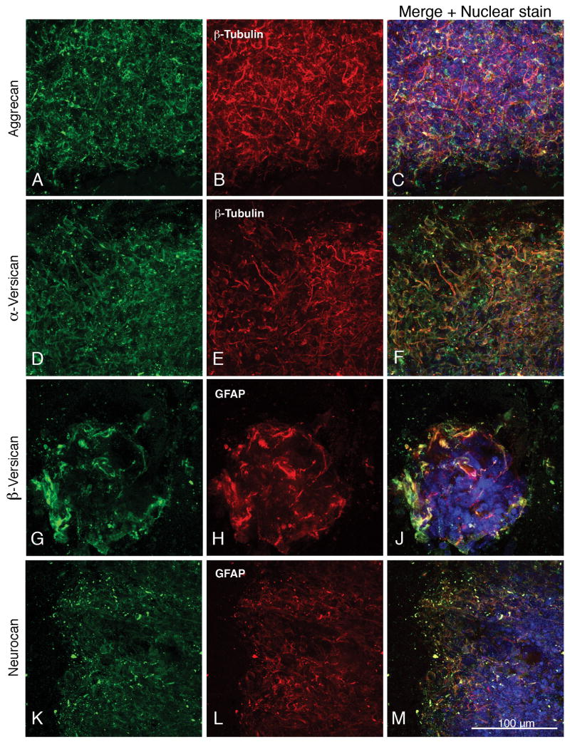
We have examined the expression and localization patterns of hyaluronan and hyaluronan-binding chondroitin sulfate proteoglycans in neural stem cells and differentiated neural cells derived from mouse embryonic stem cells. Expression of proteoglycans and hyaluronan was weak in the SSEA1-positive embryonic stem cells but increased noticeably after retinoic acid induction to nestin-positive neural stem cells. After subsequent plating, the hyaluronan-binding chondroitin sulfate proteoglycans aggrecan, neurocan, and versican are expressed by cells in both the astrocytic and neuronal lineages. During the time period that hyaluronan was present, it co-localized with each of the hyaluronan-binding proteoglycans studied and was found to be clearly associated with beta-III tubulin-expressing neurons and oligodendrocytes expressing the O4 sulfatide marker. Although proteoglycan expression levels increased to varying degrees following neural differentiation, they did not change noticably during the following 2 weeks in culture, but there was a significant decrease in hyaluronan expression. Our studies therefore demonstrate the expression by neural stem cells and neural cells derived from them of hyaluronan and its associated proteoglycans, thereby providing a necessary foundation for integrating their specific properties into developing strategies for therapeutic applications.
Figures
References
-
- Auerbach W, Dunmore JH, Fairchild-Huntress V, Fang Q, Auerbach AB, Huszar D, Joyner AL. Establishment and chimera analysis of 129/SvEv- and C57BL/6-derived mouse embryonic stem cell lines. Biotechniques. 2000;29:1024–8. 1030–1032. - PubMed
-
- Bain G, Yao M, Huettner J, Finley M, Gottlieb D. Neuronlike cells derived in culture from P19 embryonal carcinoma and embryonic stem cells. In: Banker G, Goslin K, editors. Culturing Nerve Cells. MIT; Cambridge, MA: 1998. pp. 189–212.
-
- Braam SR, Denning C, van den Brink S, Kats P, Hochstenbach R, Passier R, Mummery CL. Improved genetic manipulation of human embryonic stem cells. Nat Methods. 2008;5:389–92. - PubMed
-
- Brennan J, Skarnes WC. Gene trapping in mouse embryonic stem cells. Methods Mol Biol. 1999;97:123–38. - PubMed
Publication types
MeSH terms
Substances
Grants and funding
LinkOut - more resources
Full Text Sources
