Review
. 2010 Feb;125(2 Suppl 2):S3-23.
doi: 10.1016/j.jaci.2009.12.980.

Overview of the immune response

Affiliations
Review

Overview of the immune response

David D Chaplin. J Allergy Clin Immunol. 2010 Feb.

Abstract

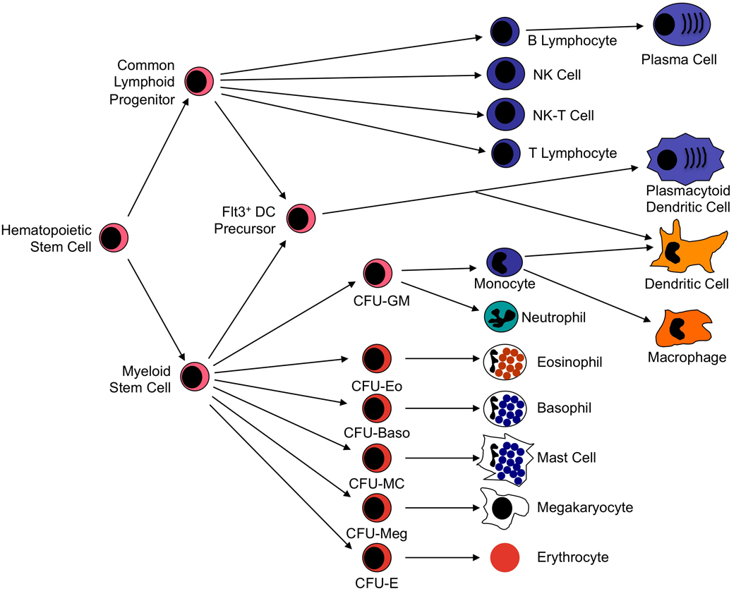
The immune system has evolved to protect the host from a universe of pathogenic microbes that are themselves constantly evolving. The immune system also helps the host eliminate toxic or allergenic substances that enter through mucosal surfaces. Central to the immune system's ability to mobilize a response to an invading pathogen, toxin, or allergen is its ability to distinguish self from nonself. The host uses both innate and adaptive mechanisms to detect and eliminate pathogenic microbes, and both of these mechanisms include self-nonself discrimination. This overview identifies key mechanisms used by the immune system to respond to invading microbes and other exogenous threats and identifies settings in which disturbed immune function exacerbates tissue injury.


Figures

Figure 1
Figure 1. Hematopoietic Stem Cell-Derived Cell Lineages
Pluripotent hematopoietic stem cells differentiate in bone marrow into common lymphoid or common myeloid progenitor cells. Lymphoid stem cells give rise to B cell, T cell, and NK cell lineages. Myeloid stem cells give rise to a second level of lineage specific colony form unit (CFU) cells that go on to produce neutrophils, monocytes, eosinophils, basophils, mast cells, megakaryocytes, and erythrocytes. Monocytes differentiate further into macrophages in peripheral tissue compartments. Dendritic cells (DC) appear to develop primarily from a DC precursor that is distinguished by its expression of the Flt3 receptor. This precursor can derive from either lymphoid or myeloid stem cells and gives rise to both classical DC and plasmacytoid DC. Classical DC can also derive from differentiation of monocytoid precursor cells. Modified with permission from Huston.
Figure 2
Figure 2. Molecular Map of the Human Major Histocompatibility Complex
The human MHC, designated HLA, is encoded on the short arm of chromosome 6. The locations of the major HLA and related genes are shown above a scale showing approximate genetic distances in kilobase pairs of DNA (kbp). The genes encoding the Class I HLA heavy chains (shown in blue) are clustered at the telomeric end of the complex. The genes encoding the Class II HLA α and β chains (shown in green) plus the genes encoding the LMP1/2, TAP1/2, and Tapascin (TAPBP) molecules (shown in orange) are clustered at the centromeric end of the complex. In between the Class I and the Class II genes are additional genes designated Class III (shown in red). These include genes encoding the cytochrome P450 21-hydroxylase (CYP21B), an inactive cytochrome P450 pseudogene (CYP21Ps), complement components C4, C2 and factor B (Bf), tumor necrosis factor (TNF), and the two lymphotoxin chains (LTA, LTB). There are two isoforms of complement C4 designated C4A and C4B. C4A interacts more efficiently with macromolecules containing free amino groups (protein antigens), whereas C4B interacts more efficiently with macromolecules containing free hydroxyl groups (glycoproteins and carbohydrates). There are genes encoding two additional HLA Class I-like molecules designated MICA and MICB (shown in purple) located between the Class III genes and the classical Class I genes. Non-functional pseudogenes are shown in gray and further designated by italics.
Figure 3
Figure 3. Structure of HLA Molecules
Molecular models derived from crystal structures of class I (A–C) and class II (D–F) HLA molecules. A, the class I α1, α2, and α3 domains are shown (light blue) in non-covalent association with the β2m molecule. Coils represent α-helices, and broad arrows represent β-strands. Anti-parallel β-strands interact to form β-sheets. The α-helices in the α1 and α2 domains form the sides and floor of a groove that binds processed antigenic peptides (yellow). The transmembrane and intracytoplasmic portions of the heavy chain are not shown. B, top view of the α1 and α2 domains displaying the antigenic peptide in a molecular complex for recognition by the TCR of a CD8+ T cell (recognition site outlined by pink rectangle). C, side view of the α1 and α2 domains highlighting the TCR contact points on both the α-helices and antigenic peptide. D, side view of the HLA class II molecule showing the α chain (light blue) and the β chain (dark blue). In the class II protein, the peptide-binding groove is made of α helices in both the α1 and β1 domains and a β-sheet formed again by both the α1 and β1 domains. E, top view of the both the α1 and β1 domains and the processed antigenic peptide fragment as they would be seen by the TCR of a CD4+ T cell. F, side view highlighting the α1 and β1 domains and the antigenic peptide. Modified with permission from Bjorkman.
Figure 4
Figure 4. Cellular Pathway for Processing and Presentation of Endogenous Antigens
Endogenous proteins are digested by the immunoproteasome to small peptide fragments. Production of the immunoproteasome is induced by IFN-γ, which leads to expression of LMP2 and LMP7 (which replace certain components of the conventional cellular proteasome) and the PA28 proteasome activator that modifies the proteasome so that it produces antigenic peptide fragments that are optimal for loading into Class I molecules. Peptides are transferred from the immunoproteasome to the endoplasmic reticulum (ER) via the TAP transporter. There the peptides are loaded, with the help of tapascin, calreticulin and the chaperone Erp57 into a class I heavy chain that associates with a β2m subunit prior to transport to the cell surface where it can be recognized by CD8+ T cells. The association of β2microglobulin with the Class I heavy chain is facilitated by an additional chaperone protein, calnexin. Modified with permission from Huston.
Figure 5
Figure 5. Cellular Pathway for Processing and Presentation of Exogenous Antigens
In the endoplasmic reticulum (ER), newly synthesized class II proteins associate with the help of calnexin with an invariant chain protein that protects the antigen-binding groove of the class II molecule until it is transported to the class II+ endosomal protein loading compartment. Exogenous antigens are taken up by phagocytosis or endocytosis, digested by the action of lysosomal enzymes, and transported to the class II+ peptide loading compartment for loading into a class II protein. There the invariant chain is proteolytically degraded and replaced by antigenic peptide with the help of the HLA-DM protein. The assembled class II protein-peptide complex is then delivered to the plasma membrane for recognition by CD4+ T cells. Modified with permission from Huston.
Figure 6
Figure 6. Differentiation and Maturation of T Cells in the Thymus
Hematopoietic stem cells which do not express CD3, CD4 or CD8 but which are committed to T cell differentiation move from the bone marrow to the thymic subcapsular zone. There they begin rearrangement of the TCR genes. Once a productive TCR β chain has been produced, they move the thymic cortex where TCR α chain rearrangement occurs and surface expression of the CD3, CD4 and CD8 proteins is induced. These CD4+CD8+ (‘double positive’) cells are positively selected on cortical epithelial cells for their ability to recognize self Class I or Class II HLA proteins. If the developing T cell has adequate affinity to recognize a self Class I protein, then it retains expression of CD8 and extinguishes expression of CD4. If the cell recognizes a self Class II protein, then it retains expression of CD4 and extinguishes expression of CD8. Selected CD4/CD8 single positive (SP) cells then move to the thymic medulla where they are negatively selected on medullary epithelial cells to remove cells with excessive affinity for self-antigens presented in HLA molecules. Cells emerge from positive selection SP for CD4 or CD8 expression and then are exported to the periphery. Cells that fail positive or negative selection are removed by apoptosis. A small fraction of cells differentiate from to rearrange their TCR γ and δ chains, rather than their TCR α and β chains. Modified with permission from Huston.
Figure 7
Figure 7. The T Cell Receptor Complex and T Cell Activation
A, the complete TCR complex includes the rearranged TCR α and β chains and also the CD3γ, CD3δ, CD3ε, and CD3ζ chains. The CD3 chains contain ITAMs in their cytoplasmic domains that can be phosphorylated to activate the intracellular signaling cascade for T cell activation. The signaling protein tyrosine kinases Lck and Fyn associate with the intracellular portions of the CD4 and CD3 chains respectively. TCR engagement by MHC plus peptide without the presence of costimulatory proteins fails to activate phosphorylation of the CD3 ITAMs and results in anergy. B, TCR engagement by MHC plus peptide with costimulatory interactions between CD28 on the T cell and CD80 or CD86 (B7.1 or B7.2) on the APC results in Lck- and Fyn-dependent phosphorylation of the CD3 chains, and recruitment of the adapter protein ZAP-70 to the CD3 complex. This leads to phosphorylation of ZAP-70, which induces the downstream program of T cell activation. C, polyclonal activation of T cells can be elicited by superantigens which interact outside the peptide binding groove with the β1 chain of the class II molecule and with all Vβ chains of a particular subclass. This activates CD4-independent, but Fyn-dependent phosphorylation of the CD3 chains, recruitment and phosphorylation of ZAP-70, and cell activation.
Figure 8
Figure 8. B Cell Differentiation and Development
B cells differentiate in the bone marrow from stem cells to become mature surface IgM and IgD expressing cells. This occurs in the absence of antigen. In peripheral lymphoid tissues, the B cell can then mature further under the influence of antigen and T cell help to undergo isotype switching and affinity maturation by somatic mutation. The factors controlling the final differentiation from antibody-secreting B cell to plasma cell are incompletely characterized, but require the participation of the transcription factors Blimp1, Xbp1 and IRF4. Correlations are show between the stage of cell differentiation and the expression of key molecules in the cell (TdT, RAG1/RAG2, cytoplasmic μ) and on the cell surface (class II, CD19, CD21, CD25, CD45, and surface Ig). Modified with permission from Huston.
Figure 9
Figure 9. The Activation Pathways of Complement
Three pathways lead to activation of complement. The Classical Pathway is initiated by complexes of IgM, IgG1, or IgG3 with antigens. This activates proteolysis of C1 that cleaves C4 and C2 to form the classical pathway C3 convertase. The Mannose Lectin Pathway is activated by interaction of mannan-containing microbes with MBL, which activates MASP-1 and MASP-2 to cleave C4 and C2, again forming a C3 convertase. The Alternative Pathway is initiated by interactions between microbial antigens and inhibitory complement regulatory proteins. This permits autoactivation of the pathway in which C3 interacts with factor B and factor D to generate the alternative pathway C3 convertase. These convertases all cleave C3 to generate the anaphylatoxic C3a fragment and depositing C3b on the activating microbial particle or immune complex. This opsonizes the particle for phagocytosis and initiates the activation of the membrane attack complex. The C5a fragment that is proteolytically released from C5 also is a highly anaphylatoxic molecule that induces intense local inflammation.

References

    1. Hiemstra PS. The role of epithelial beta-defensins and cathelicidins in host defense of the lung. Exp Lung Res. 2007;33:537–542. - PubMed
    1. Holmskov U, Thiel S, Jensenius JC. Collectins and ficolins: humoral lectins of the innate immune defense. Annu Rev Immunol. 2003;21:547–578. - PubMed
    1. Sjoberg AP, Trouw LA, Blom AM. Complement activation and inhibition: a delicate balance. Trends Immunol. 2009;30:83–90. - PubMed
    1. Robin C, Bollerot K, Mendes S, Haak E, Crisan M, Cerisoli F, et al. Human placenta is a potent hematopoietic niche containing hematopoietic stem and progenitor cells throughout development. Cell Stem Cell. 2009;5:385–395. - PMC - PubMed
    1. Jonsson AH, Yokoyama WM. Natural killer cell tolerance licensing and other mechanisms. Adv Immunol. 2009;101:27–79. - PubMed