

Lack of association between 11C-PiB and longitudinal brain atrophy in non-demented older individuals
- PMID: 20176414
- PMCID: PMC2908199
- DOI: 10.1016/j.neurobiolaging.2009.12.008
Lack of association between 11C-PiB and longitudinal brain atrophy in non-demented older individuals
Abstract
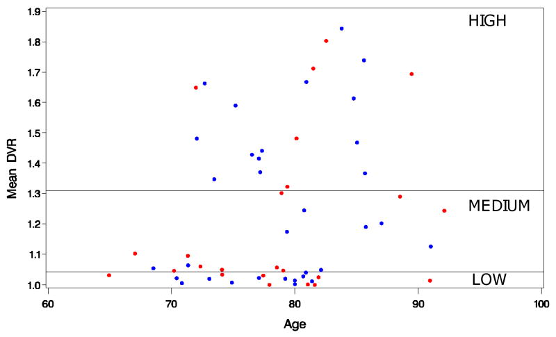
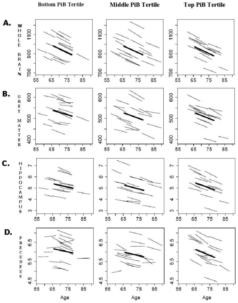
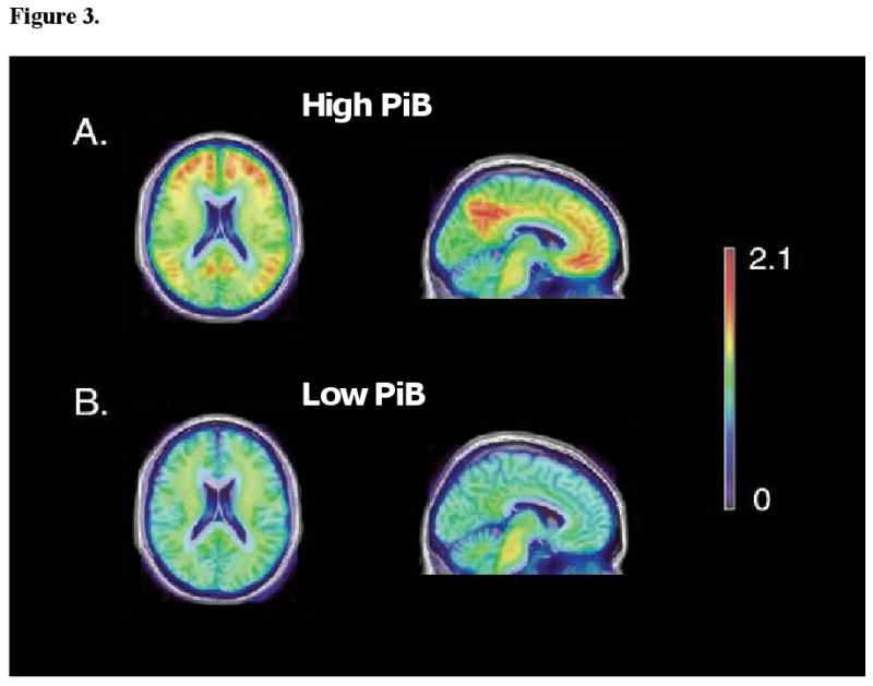
Amyloid-β plaques (Aβ) are a hallmark of Alzheimer's disease (AD), begin deposition decades before the incipient disease, and are thought to be associated with neuronal loss, brain atrophy and cognitive impairment. We examine associations between (11)C-PiB-PET measurement of Aβ burden and brain volume changes in the preceding years in 57 non-demented individuals (age 64-86; M=78.7). Participants were prospectively followed through the Baltimore Longitudinal Study of Aging, with up to 10 consecutive MRI scans (M=8.1) and an (11)C-PiB scan approximately 10 years after the initial MRI. Linear mixed effects models were used to determine whether mean cortical (11)C-PiB distribution volume ratios, estimated by fitting a reference tissue model to the measured time activity curves, were associated with longitudinal regional brain volume changes of the whole brain, ventricular CSF, frontal, temporal, parietal, and occipital white and gray matter, the hippocampus, orbito-frontal cortex, and the precuneus. Despite significant longitudinal declines in the volumes of all investigated regions (p<0.05), no associations were detected between current Aβ burden and regional brain volume decline trajectories in the preceding years, nor did the regional volume trajectories differ between those with highest and lowest Aβ burden. Consistent with a threshold model of disease, our findings suggest that Aβ load does not seem to affect brain volume changes in individuals without dementia.
Published by Elsevier Inc.
Conflict of interest statement
Ira Driscoll - no actual or potential conflicts of interest.
Yun Zhou - no actual or potential conflicts of interest.
Yang An - no actual or potential conflicts of interest.
Jitka Sojkova - no actual or potential conflicts of interest.
Christos Davatzikos - no actual or potential conflicts of interest.
Michael A Kraut - no actual or potential conflicts of interest.
Weiguo Ye - no actual or potential conflicts of interest.
Luigi Ferrucci - no actual or potential conflicts of interest.
Chester A Mathis - GE Healthcare holds a license agreement with the University of Pittsburgh based on the PiB technology described in this manuscript. Dr. Mathis is a co-inventor of PiB and, as such, has a financial interest in this license agreement.
William E Klunk - GE Healthcare holds a license agreement with the University of Pittsburgh based on the PiB technology described in this manuscript. Dr. Klunk is a co-inventor of PiB and, as such, has a financial interest in this license agreement.
Dean F Wong - no actual or potential conflicts of interest.
Susan M Resnick – no actual or potential conflicts of interest.
Figures
References
-
- Alzheimer A. Über einen eigenartigen schweren Krankheitsprozess der Hirnrinde. Neurologisches Centralblatt. 1906;25:1134.
-
- Alzheimer A. Über eine eigenartige Erkrankung der Hirnrinde. Allgemeine Zeitschrift für Psychiatrie und psychisch-gerchtliche Medizin. 1907;64:146–148.
-
- Bennett DA, Schneider JA, Arvanitakis Z, Kelly JF, Aggarwal NT, Shah RC, Wilson RS. Neuropathology of older persons without cognitive impairment from two community-based studies. Neurology. 2006;66:1837–1844. - PubMed
-
- Blessed G, Tomlinson BE, Roth M. The association between quantitative measures of dementia and of senile change in the cerebral grey matter of elderly subjects. Br J Psychiatry. 1968;114:797–811. - PubMed
-
- Braak H, Braak E. Neuropathological staging of Alzheimer-related changes. Acta Neuropathol. 1991;82:239–259. - PubMed
Publication types
MeSH terms
Substances
Grants and funding
LinkOut - more resources
Full Text Sources
