Review
. 2010 May;18(5):215-23.
doi: 10.1016/j.tim.2010.01.001. Epub 2010 Feb 20.

Structural evaluation of new human polyomaviruses provides clues to pathobiology

Affiliations
Review

Structural evaluation of new human polyomaviruses provides clues to pathobiology

Edward M Johnson. Trends Microbiol. 2010 May.

Abstract

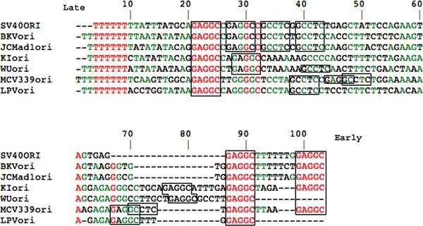
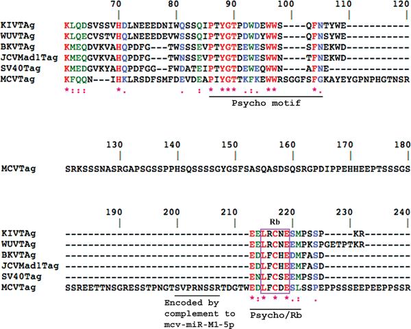
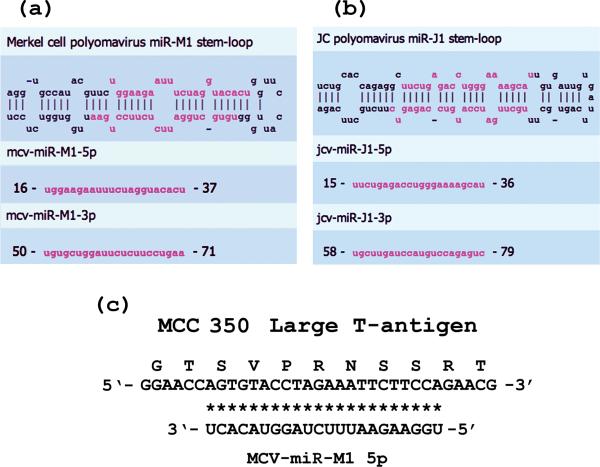
In the past three years, remarkable discoveries have added three new human polyomaviruses (KI virus (KIV), WU virus (WUV) and Merkel cell virus (MCV)) to a class that previously had only two disease-causing members (BK virus (BKV) and JC virus (JCV)) identified. Two monkey polyomaviruses, simian virus (SV)40 and B-cell lymphotropic polyomavirus (LPV) are also present in humans. KIV and WUV lack the agnoprotein coding sequence and regulatory micro (mi)RNA clusters of BKV, JCV and SV40. MCV lacks the agnoprotein sequence but generates miRNAs. KIV, WUV and MCV are all widespread in humans. Although they have distinctive tissue tropisms, all these viruses are probably acquired in childhood. Of these viruses, only MCV has thus far been strongly linked to cancer. Marshalled evidence from diverse sources implicates MCV as an etiological agent of Merkel cell carcinoma. This review compares the structural features of the new and previously known polyomaviruses, with the aim of identifying approaches to molecular pathology.


Figures

Figure 1
Figure 1. Distinct sequence arrangements in origins of DNA replication of newly discovered human polyomaviruses
DNA sequences, shown 5’ to 3’ for one strand, were compared for origins of DNA replication of the known human polyomaviruses and for Simian virus 40 (SV40) and Monkey B-cell lymphotropic polyomavirus (LPV) using Multalin [71]. SV40 is included in the figure because of its presence in humans. LPV is included because many humans are reportedly seropositive for it or a homologous virus [65]. Sequences were obtained as described in Box 1. Red lettering denotes 100% identity among all the origins; green lettering denotes >50% identity. Numbering is from the first residue shown for MCV. T-antigen-binding elements, GAGGC or the complement GCCTC, are boxed. MCV is the only virus shown to have the overlapping, palindromic element GAGGCCTC. MCV has two of these elements opposing each other enclosing a highly A-rich segment. These two unusual elements[16] could affect binding of T-antigen hexamers and initiation of DNA replication.
Figure 2
Figure 2. Protein sequence alignment of Rb-binding regions of large T-antigens of human polyomaviruses
The alignment employed Clustal W [72], which weighs conservative substitutions in aas. Numbering begins based on the SV40 T-antigen sequence. 100% identities are in red lettering and denoted below by “*”. Green lettering and a “:” denote highly conserved aa's. Blue lettering and “.” denote a less conservative aligned aa. The motif LXCXE (boxed) is essential in T-antigens for binding the retinoblastoma tumor suppressor protein, Rb. The Psycho motif is conserved among T-antigens and influences Rb activities. Note that only in MCV is this motif interrupted by a sequence extending from aa 106 to aa 212. The central line of sequence from 121 to 180 is shown for MCV alone because none of the other T-antigens have any sequence in this region. This interrupting sequence could represent an intron, but there are no stop codons preventing its translation. It contains a sequence, SVPRNSSR, encoded by a perfect complement to the miRNA mcv-miR-M1-5p (Figure 3). This sequence in MCV almost immediately precedes the Rb-binding LFCDE motif. LPV is not included in Figure 2 because, except for certain of the important motifs, it is not overtly homologous to several of the other T-antigens.
Figure 3
Figure 3. MiRNAs and pre-miRNAs generated by MCV and JCV
MiRNAs for MCV [37] and JCV [36] have recently been reported. Alignment of stem-and-loop structures for an MCV pre-miRNA (a) and a JCV pre-miRNA (b) are shown and have been adapted from the mirbase website (www.mirbase.org). Sequences of the 22 nt miRNAs are shown in pink lettering. A perfect match between mcv-miR-M1-5p and a DNA sequence encoding a segment of MCV large T-antigen is shown in (c). As shown in Figure 2, this sequence is N-terminal to the Rb-binding motif of MCV T-antigen.

Similar articles

Cited by

References

    1. Allander T, Andreasson K, Gupta S, Bjerkner A, Bogdanovic G, Persson MA, Dalianis T, Ramqvist T, Andersson B. Identification of a third human polyomavirus. J Virol. 2007;81:4130–4136. - PMC - PubMed
    1. Gaynor AM, Nissen MD, Whiley DM, Mackay IM, Lambert SB, Wu G, Brennan DC, Storch GA, Sloots TP, Wang D. Identification of a novel polyomavirus from patients with acute respiratory tract infections. PLoS Pathog. 2007;3:e64. - PMC - PubMed
    1. Feng H, Shuda M, Chang Y, Moore PS. Clonal integration of a polyomavirus in human Merkel cell carcinoma. Science. 2008;319:1096–1100. - PMC - PubMed
    1. Hogan TF, Borden EC, McBain JA, Padgett BL, Walker DL. Human polyomavirus infections with JC virus and BK virus in renal transplant patients. Ann Intern Med. 1980;92:373–378. - PubMed
    1. Trofe J, Hirsch HH, Ramos E. Polyomavirus-associated nephropathy: update of clinical management in kidney transplant patients. Transpl Infect Dis. 2006;8:76–85. - PubMed