. 2010 Jul;27(7):1645-58.
doi: 10.1093/molbev/msq052. Epub 2010 Feb 22.

Evolutionary dynamics of the wnt gene family: a lophotrochozoan perspective

Affiliations

Evolutionary dynamics of the wnt gene family: a lophotrochozoan perspective

Sung-Jin Cho et al. Mol Biol Evol. 2010 Jul.

Abstract

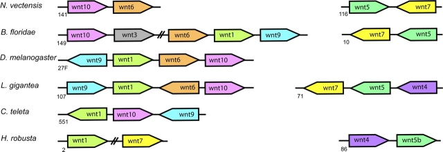
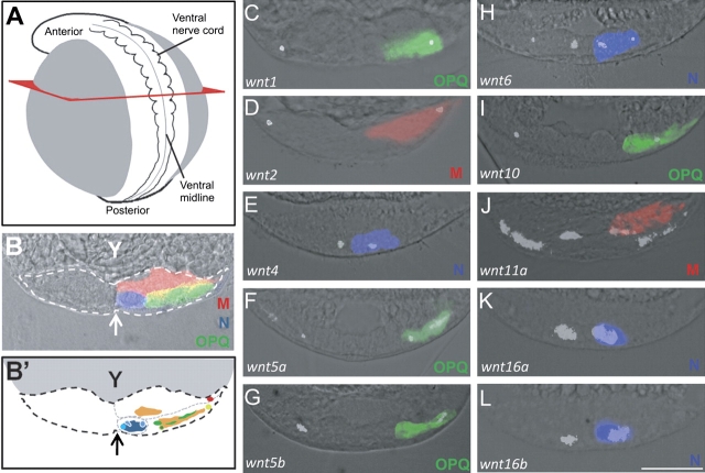
The wnt gene family encodes a set of secreted glycoproteins involved in key developmental processes, including cell fate specification and regulation of posterior growth (Cadigan KM, Nusse R. 1997. Wnt signaling: a common theme in animal development. Genes Dev. 11:3286-3305.; Martin BL, Kimelman D. 2009. Wnt signaling and the evolution of embryonic posterior development. Curr Biol. 19:R215-R219.). As for many other gene families, evidence for expansion and/or contraction of the wnt family is available from deuterostomes (e.g., echinoderms and vertebrates [Nusse R, Varmus HE. 1992. Wnt genes. Cell. 69:1073-1087.; Schubert M, Holland LZ, Holland ND, Jacobs DK. 2000. A phylogenetic tree of the Wnt genes based on all available full-length sequences, including five from the cephalochordate amphioxus. Mol Biol Evol. 17:1896-1903.; Croce JC, Wu SY, Byrum C, Xu R, Duloquin L, Wikramanayake AH, Gache C, McClay DR. 2006. A genome-wide survey of the evolutionarily conserved Wnt pathways in the sea urchin Strongylocentrotus purpuratus. Dev Biol. 300:121-131.]) and ecdysozoans (e.g., arthropods and nematodes [Eisenmann DM. 2005. Wnt signaling. WormBook. 1-17.; Bolognesi R, Farzana L, Fischer TD, Brown SJ. 2008. Multiple Wnt genes are required for segmentation in the short-germ embryo of Tribolium castaneum. Curr Biol. 18:1624-1629.]), but little is known from the third major bilaterian group, the lophotrochozoans (e.g., mollusks and annelids [Prud'homme B, Lartillot N, Balavoine G, Adoutte A, Vervoort M. 2002. Phylogenetic analysis of the Wnt gene family. Insights from lophotrochozoan members. Curr Biol. 12:1395.]). To obtain a more comprehensive scenario of the evolutionary dynamics of this gene family, we exhaustively mined wnt gene sequences from the whole genome assemblies of a mollusk (Lottia gigantea) and two annelids (Capitella teleta and Helobdella robusta) and examined them by phylogenetic, genetic linkage, intron-exon structure, and embryonic expression analyses. The 36 wnt genes obtained represent 11, 12, and 9 distinct wnt subfamilies in Lottia, Capitella, and Helobdella, respectively. Thus, two of the three analyzed lophotrochozoan genomes retained an almost complete ancestral complement of wnt genes emphasizing the importance and complexity of this gene family across metazoans. The genome of the leech Helobdella reflects significantly more dynamism than those of Lottia and Capitella, as judged by gene duplications and losses, branch length, and changes in genetic linkage. Finally, we performed a detailed expression analysis for all the Helobdella wnt genes during embryonic development. We find that, although the patterns show substantial overlap during early cleavage stages, each wnt gene has a unique expression pattern in the germinal plate and during tissue morphogenesis. Comparisons of the embryonic expression patterns of the duplicated wnt genes in Helobdella with their orthologs in Capitella reveal extensive regulatory diversification of the duplicated leech wnt genes.


Figures

F<sc>IG</sc>. 1.
FIG. 1.
ML tree showing the distribution of the wnt genes. Numbers on branches are pp/bs values. Species abbreviations: Api, Acyrthosiphon pisum; Ate, Achaearanea tepidariorum; Bfl, Branchiostoma floridae; Cte, Capitella teleta; Hro, Helobdella robusta; Hsa, Homo sapiens; Lgi, Lottia gigantea; Nve, Nematostella vectensis; Spu, Strongylocentrotus purpuratus; Tca, Tribolium castaneum; Xtr, Xenopus tropicalis. Lophotrochozoan genes are in bold. Black stars indicate the lineages tested for positive selection (for details, see Supplementary Material, Supplementary Material online).
F<sc>IG</sc>. 2.
FIG. 2.
wnt genetic linkage shared among metazoans. Cartoon showing the relative position and orientation of the wnt genes that share scaffolds/chromosome in Nematostella vectensis, Branchiostoma floridae, Drosophila melanogaster, Lottia gigantea, Capitella teleta, and Helobdella robusta. The distances between genes and the size of the wnt genes themselves are not to scale, but slanting lines indicate very large intergenic regions. Numbers indicate the specific scaffold or chromosome (in the case of D. melanogaster).
F<sc>IG</sc>. 3.
FIG. 3.
Expression patterns of the nonduplicated leech wnt genes during embryogenesis. Leech development from zygote to juvenile has been divided into 11 stages; schematic views of selected stages are shown in the bottom row (vnc, ventral nerve cord; fg, foregut; mg, midgut). Columns I–IV show nitro blue tetrazolium chloride/5-bromo-4-chloro-3-indolyl phosphate coloration reactions and columns V–VII show FISH. Columns I and II illustrate animal views at two-cell stage at earlier (I; 290–310 min AZD) and later (II; 320–340 min AZD) time points. Two main patterns are observed, either a constant and stable association of the mRNA with the teloplasm in cell CD throughout the two-cell stage (i.e., wnt2 and wnt6) or diffuse and dynamic expression, with mRNA accumulating first in CD (295–310 min AZD) and then in AB (320–340 min AZD; i.e., wnt1 and wnt7). Column III depicts animal views of embryos at stage 4. At this stage, there are four macromeres and 4–15 micromeres, depending on the exact development time. In all cases except for wnt4, the mRNA is present in the nucleoplasm (i.e., wnt5a and wnt16a) of the four macromeres and in most cases in teloplasm (i.e., wnt7 and wnt10). wnt4, however, is found exclusively in the nuclei of the micromeres. Column IV shows animal views of stage 7 embryos. At this point, the embryo contains ten teloblasts, each of which gives rise to a column (bandlet) of blast cells, whose progeny will generate the segmented ectoderm and mesoderm; the progeny of the micromeres, which will form nonsegmental body parts (i.e., proboscis); and the A′′′, B′′′, and C′′′ macromeres, which will contribute to the midgut. Most wnt genes at this stage are strongly expressed in both teloblasts and bandlets (except for wnt11b which is absent and wnt7 which shows weak expression in the bandlets). In addition, wnt6, wnt11a, and wnt16a are expressed in the micromere-derived epithelium. Columns V (lateral view) and VI (ventral view showing details of the expression patterns in the germinal plate) illustrate stage 9 embryos. Column VI, FISH where red shows the wnt mRNAs. All wnt genes except wnt7 and wnt11b are expressed in segmentally iterated patterns in the germinal plate. Additionally, wnt7 and wnt16a are expressed in the earliest stages of proboscis development. Column VII shows lateral views of stage 10 embryos, except for wnt1, wnt5a, wnt7, and wnt11b, which now appear in the developing proboscis, every other wnt gene is expressed in a pattern similar to that observed in stage 9. Scale bar, columns I through V and VII, 100 μm; column VI, 50 μm.
F<sc>IG</sc>. 4.
FIG. 4.
Expression patterns of the duplicated leech wnt genes during embryogenesis. Details as described for figure 3.
F<sc>IG</sc>. 5.
FIG. 5.
Germ layer localization of wnt-expressing cells in Helobdella germinal plate. (A) Drawing of a stage 9 embryo showing the germinal plate and ventral nerve cord (white) and underlying yolk (gray). The red square shows where the cross sections were done. (B) Pseudocolored image (combined brightfield and fluorescence imaging) showing yolk (Y) and germinal plate (dotted contour) in a cross section of a stage 9 embryo in which the ventral (N) and dorsolateral (OPQ) ectodermal lineages on the right side of the germinal plate are marked with FRDA (blue) and FDA (green), respectively, and the mesodermal (M) lineage is marked with RDA (red), by lineage tracer injection into the various precursor cells at stage 6a (for details, see Materials and Methods). Arrow indicates ventral midline. (B′) Schematic representation of (B), summarizing the expression of the data shown in (CL); wnt1, olive green; wnt2, red; wnt4, light blue; wnt5a and wnt5b, light green; wnt6, medium blue; wnt10, dark green; wnt11a, orange; wnt16a and wnt16b, dark blue. The apparent overlap in this transverse view between wnt5a and wnt5b and between wnt16a and wnt16b is resolved in ventral views shown in figure 6. (CL) Cross sections of stage 9 embryos similar to that shown in panel B, but in which only one lineage was labeled, and which had been processed by FISH for the indicated wnt genes (light gray). Scale bar, 100 μm.
F<sc>IG</sc>. 6.
FIG. 6.
Expression patterns of the leech-duplicated wnt genes in late development stages. Pseudocolored confocal stacks of DFISH-processed embryos showing ventral views of segmental tissues at stage 9 (A, C, E) and lateral views of the proboscis, counterstained with 4′,6-diamidino-2-phenylindole to reveal nuclei (blue) at stage 10 (B, D, F). (A and B) wnt5a (red) and wnt5b (green) are both expressed in midbody segments, with little or no overlap; only the wnt5a paralog is expressed in the proboscis. (C) wnt11a (red) and wnt11c (green) are expressed in partially overlapping patterns within the caudal segments. (D) Within the proboscis, wnt11a (red) and wnt11b (green) are expressed in discrete sets of posterior and anterior cells, respectively. (E and F) wnt16a (red) and wnt16b (green) are both expressed in neighboring sets of cells with little or no overlap in both midbody segments and at the anterior tip of the proboscis. Scale bars: A, C, and E 100 μm; B, D, and F 50 μm.
F<sc>IG</sc>. 7.
FIG. 7.
Expression of wnt5, wnt11 and wnt16 genes in Capitella. Whole-mount ISH of stage 7 Capitella larvae showing expression domains of wnt5 (AD), wnt11 (EH), and wnt16 (IL). All panels are anterior to the left. A, E, and I are lateral views, with ventral down; all other panels are ventral views. BD, FH, and JL are series of distinct focal planes, progressing dorsally from the ventral-most plane (B, F, J). BD are the same animal and KL are the same animal. I is a merge of multiple focal planes. Brackets mark the ventral nerve cord, and the mouth is marked with an asterisk, except in J, so as not to obscure the in situ signal. Br, brain; fg, foregut; hg, hindgut; mes, mesoderm; pgz, posterior growth zone; vnc, ventral nerve cord. Scale bar, 50 μm.

References

    1. Abascal F, Zardoya R, Posada D. ProtTest: selection of best-fit models of protein evolution. Bioinformatics. 2005;21:2104–2105. - PubMed
    1. Blake JA, Grassle JP, Eckelbarger KJ. Capitella teleta, a new species designation for the opportunistic and experimental capitellid, Capitella sp. I, with a review of the literature for confirmed records. Zoosymposia. 2009;2:25–53.
    1. Bolognesi R, Farzana L, Fischer TD, Brown SJ. Multiple Wnt genes are required for segmentation in the short-germ embryo of Tribolium castaneum. Curr Biol. 2008;18:1624–1629. - PMC - PubMed
    1. Burch JB. Regulation of GATA gene expression during vertebrate development. Semin Cell Dev Biol. 2005;16:71–81. - PubMed
    1. Cadigan KM, Nusse R. Wnt signaling: a common theme in animal development. Genes Dev. 1997;11:3286–3305. - PubMed

Publication types